Anemias-Fe deficiency Flashcards
What is anemia?
What factors affect wheater a cetain mass of red cells is adeaquate?
Anemia is defined as insufficient red cell mass to adequately deliver oxygen to peripheral tissues.
The factors influencing whether a certain red cell mass is adequate include the oxygen carrying capacity of the red cells (depends on Hgb) and metabolic demand of the tissues.
For example a patient with cyanotic congenital heart disease (decreased hemoglobin oxygen saturation) may have an equal red cell mass as a patient with hypothyroidism (decreased metabolic demands), but the patient with heart disease may be anemic whereas the hypothyroid patient may be not given the balance of oxygen delivery to the tissues.
What blood parameters are used to determine is anemia is present?
What is defined as anemia in terms of these parameters?
Whats special about infants at birth in terms of hemoglobing and hematocrit concentration?
What about in childhood?
What about menstruting women?
Measurements of ______ are used to determine is anemia is present:
1- hemoglobin concentration (Hgb, g/dL)
2- hematocrit (Hct, %)
3- red blood cell count (cells x 10x12/L)
- We define anemia as a Hgb, Hct or RBC count less than 2 standard deviations below the mean (< 2.5th percentile) for age, gender and ethnicity.
- The table attached provides an illustration of the typical variation in hemoglobins based on age and gender.
At birth, infants have a very high hemoglobin concentration and hematocrit which in the first 8 weeks decreases to lower levels.
During childhood, the levels of Hgb and Hct are lower than adults until the onset of puberty after which the values reach adult levels.
**Menstruating women have lower Hgb and Hct values than men, in part, because of more tenuous iron stores.

What routine laboratory measurements are used to define anemia clinically?
What measurement is helpful in determining rate of production of RBCs?
To define anemia clinically we include the:
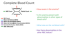
- Complete blood count: composed of Hgb, Hct, RBC count, the mean corpuscular volume or MCV (average volume of red blood cells), the mean corpuscular hemoglobin concentration or MCHC (mean corpuscular Hb concnetration), the red cell distribution width or RDW (varition in size of the peripheral blood RBCs), the white blood cell count and a differential consisting of the various types of white cells (as %), and a platelet count.
- Valuable information can be obtained by reviewing a blood smear stained with Wright’s stain to observe any changes in red cell morphology.
- In addition, the reticulocyte count or reticulocyte production index (RPI) is helpful to determine the rate of production.
How can you identify reticulocytes?
What is the normal range for reticulocytes?
What is the reticulocyte index (RI)?
- Reticulocytes can be identified by the presence of mRNA in the cells for their first day in the circulation. These can be identified by the darker appearance of these cells on a routine peripheral smear or by staining of the mRNA with certain supra-vital dyes.
- Reticulocytes are formally determined as the percent of 1,000 red cells counted and the normal range is from 0.4-1.7%. Increased red cell production is associated with a 3.5 to 5-fold (6-8 fold during anemia) increase over this baseline normal range. The calculation of the absolute reticulocyte count (which is equal to the percent of reticulocytes times the RBC count) is helpful in determining the relevance of the reticulocyte count; anything over 50,000/mcl is considered an increase over baseline maintenance production of red cells.
Reticulocyte index (RI) is another measurement of the production of red cells and is a way to correct the reticulocyte count for red cell concentration and stress reticulocytosis. The corrected reticulocyte count or the reticulocyte index provides a ratio of how many fold beyond baseline the production of red cells is.
What is the stress factor (correction factor) and how it is correlated with RI?
Where the stress factor = 1.5 (mild anemia > 9 g/dL)
- 0 (No anemia >12 g/dL)
- 0 (moderate anemia 6.5-9)
- 5 (severe anemia < 6.5)
The reticulocyte index should be between 1.0 and 2.0 for a healthy individual.
A RI < 2 with anemia indicates decreased production of reticulocytes and therefore red blood cells.
A RI > 3 with anemia indicates loss of red blood cells (destruction, bleeding, etc) leading to increased compensatory production of reticulocytes to replace the lost red blood cells.

Describe the different ways in which the complete blood count aids in anemia parameters?
How is the time over which anemia develops important in its presentation?
What are the symptoms of anemia?
The time over which the anemia develops also plays a role in the clinical presentation of the patient with anemia.
- In anemias which develop over weeks, 2,3-DPG within the cells will increase making the dissociation of oxygen to the tissues more efficient to compensate for the low oxygen carrying capacity.
- If anemia develops acutely (hours-days), there is not enough time to establish this compensatory mechanism.
Symptoms of anemia include shortness of breath (tightness of the chest, hunger for air, feeling of suffocation) , fatigue, headache, dizziness, claudication or pain with exercise, or pallor.
Sample of peripheral blood smear and describe what the terms mean:

Microcytes: an unusually small red blood cell (MCV<80)
Macrocytes: larger than usual red blood cells (MCV >100)
Spherocyte: red blood cells (RBCs)) or erythrocytes that are sphere-shaped rather than bi-concave disk shaped. Spherocytes are found in all hemolytic anemias to some degree.
Target cells: RBC cells with “bulls-eye appearence”
Schistocyte: schizocyte (from Greek schistos for “divided” or schistein for “to split”, and kytos for “hollow” or “cell”) is a fragmented part of a red blood cell. Schistocytes are typically irregularly shaped, jagged, and have two pointed ends.
What is the difference in the presentation of chronic vs. acute anemias?
If the anemia is mild or chronic and slowly progressive, the vital signs may be normal.
If the anemia is _severe or sudden onse_t, the patient may be tachycardic (fast heart rate) or hypotensive (low blood pressure). A heart murmur (sloshing sound) can often be heard if the anemia is severe.
What are the questions to ask when classifying anemia:
1- Are there any additional hematologic abnormalities besides anemia (e.g. low platelet or white blood cell count)? If the answer is yes, then search for infiltrative and proliferative processes (e.g. leukemia, lymphoma, aplastic anemia).
2- If the only manifestation is anemia, the reticulocyte count is examined. If there is an increase in reticulocytes, then one considers the possibility of increased red blood cell destruction (hemolysis) or perhaps the possibility of blood loss and response to hemorrhage.
3- If the reticulocyte count is not increased and there is no other evidence for hemolysis, then one can consider the type of anemia based on the MCV and size, i.e. normocytic, macrocytic (large RBCs) or microcytic (small) RBCs.

Iron deficiency is a type of underproduction anemias characterized by what type of RBC?
Iron deficiency is the most common cause of anemia and is estimated to affect more than 1 billion people globally. This is the prototypic microcytic anemia (<80).
Iron in the body exists in what two states? Which one binds to Hb?
What happens to iron in aqueous solutions?
When is iron soluble?
Iron balance is solely regulated by what process?
- Iron exists in two valence states (ferric, Fe3+ or ferrous, Fe2+) and activity may depend on a specific state. Ferrous Iron binds to Hgb.
- In aqueous solution iron forms insoluble iron hydroxides unless bound to a chaperone protein
- Iron is more soluble at low pH
- Iron losses are fixed (exfoliation of skin, GI mucosa, menstruation) so iron balance is controlled by regulation of absorption. Why? As there is no active mechanism for regulating excretion from the body.
Explain the difference between the two iron protein chaperones:

How is iron distributed inthe body?
Where is the majority of the Iron contained?
Iron is distributed throughout the body in many different forms.
- The majority of iron is contained in hemoglobin (65%).
- About 6% of total body iron is in myoglobin which relates to muscle oxygen storage.
- Ferritin (soluble iron-binding protein that stores iron in macrophages and hepatocytes, serum levels directly correlate with bone marrow iron stores, so if a decrease in iron occurs, a decrease in ferritin will occur) and hemosiderin (insoluble product of ferritin degradation in lysosomes, does not circulte in serum) are the primary storage forms of iron (25% of the total body iron, mostly intracellular). Ferritin is able to bind up to 4,500 atoms of iron and is available to replenish other iron containing compounds throughout the body.
A very small amount of iron is bound to transferrin (1%), the transport protein which moves the iron to tissues requiring iron, particularly the developing erythroid precursors.
The remainder of iron (<3-4%) is associated with a whole variety of enzymes, including catalases, peroxidases, cytochromes, and other proteins which are critical to basic metabolic processes of the cell.

Regulation of iron hemostasis, dietary iron of present in what two form? Which one is absorbed more efficiently?
Iron homeostasis is primarily regulated via adjustments to absorption of dietary iron since iron losses are fixed.
Dietary iron is present in two forms:
1-heme-iron (usually animal-derived) and 2) elemental or non-heme iron.
-Heme-iron is absorbed much more efficiently than elemental iron via a mechanism which is unclear.
Where is elemental Iron absorbed?
What are the steps? Mention the name of the transporters and enzymes:
What transporter transports Fe out of the cell on the basolateral membrane?
What is the name of the Iron oxidase in the plasma that changes ferrous to ferric iron?
Elemental iron is absorbed in the proximal duodenum.
Steps:
1) Ferric iron is reduced to ferrous iron at the _apical brush border i_n the lumen of the duodenum by an iron-reductase believed to be duodenal cytochrome B (DcytB).
2- Once reduced, the ferrous iron is transported into the enterocyte by divalent metal transporter 1 (DMT1).
3- It can then be stored as ferritin or exported into the plasma by ferroportin, an iron transporter on the baso-lateral membrane.
4- Once in the plasma, the ferrous iron is oxidized by an iron-oxidase (likely hephaestin) and then bound to transferrin to be transported to the bone marrow where it will be utilized for erythropoiesis.

What is Hepcidin and what is its importance in iron metabolism?
Where is it synthesized? Excreted?
When does itc concentration increase? Decrease?
It is a 25 amino acid peptide hormone.
- Synthesized in the liver, circulates in plasma, excreted by kidneys.
- It is a Negative regulator of cellular iron export through down regulation of ferroportin, can bind to it and cause its degradation.
Thus:
It increases the concentration of iron in ferritin (inside cell).
- Inhibits duodenal iron absorption into the plasma
- Inhibits macrophage iron release
- Increases in response to infection, inflammation, iron overload
- Decreases in response to hypoxia, iron deficiency

The Iron cycle:
Transferrin bind Fe in what valence state?
Where does transferrin brings Fe to?
Once in the bone marrow how does transferrin transfers its Iron?
Once through the mucosal cell and bound to transferrin, iron enters the iron cycle.
Transferrin is the main transport protein for iron. An 84 kDa plasma protein produced in the liver which binds two moles of iron in the ferric form for each mole of protein, the binding specificity and affinity for iron by transferrin are very high and stable, even at decreaed pH.
Transferrin, which under normal conditions, is partially saturated with iron, finds its way to the bone marrow and the maturing normoblasts.
Process:
1- Transferrin binds *transferrin receptors on the surface of the normoblasts.
The transferrin receptor provides direction for the iron cycle. Iron bound to plasma transferrin is delivered to the developing normoblast through interaction with the transferrin receptor. The transferrin/transferrin receptor complex enters the cell through an invagination of clathrin coated pits to form specialized endosomes. These endosomes become acidified through the entry of protons releasing iron from transferrin. The iron exits the endosome through DMT1 to go to sites of storage (i.e. ferritin within the cell) and the transferrin receptor/transferrin complex returns to the cell surface where it is externalized and apotransferrin is released, free to pick up iron for the next cycle.
2- As erythrocytes are produced, they are released into the circulating pool and, after 120 days, are removed from the circulation by macrophages in the spleen.
3- Once intracellular, macrophages turn over the cells and sequester the iron in ferritin stores. These molecules have a similar structure and are mainly found in an intracellular location. Small amounts are extravascular and their concentration is under steady state conditions proportional to intracellular levels.

What is the importance of the iron cycle?

The importance of the iron cycle is that it allows most of the recyclable (red cell) iron to be re-used and minimizes the amount of iron required from absorption each day.
What are the causes of Iron deficiency?
•Decreased iron intake
–>Insufficient dietary intake (exclusively breastfed babies, anorexics, vegans)
•Decreased iron absorption (excessive intake of absorption inhibitors, inflammatory bowel disease)
•Prematurity
•Increased iron losses
•Excessive blood loss (GI tract, menstruation, lungs)
•Increased iron utilization
•Growth
•Pregnancy

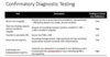
Parameters of comfirmatory testing for Iron deficiency:
Progression of iron deficiency:
