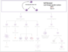
Hematopoiesis Flashcards
What is hematopoiesis?
Where does it occur in adults?
Hematopoiesis is the complex process that results in the formation of the mature, functional red blood cells, white blood cells, and platelets in the peripheral blood, and ultimately the functional cells of the lymphoid organs, and reticuloendothelial system.
In the normal adult, this process occurs exclusively in the bone marrow.
Pre‐natal hematopoiesis
Where are RBCs produces up to the third month of gestation?
What about fromt he second to the seventh month of gestation?
1- Primitive blood cells, primarily red cells are first produced in the yolk sac. Hematopoiesis in the yolk sac is largely finished by the third month of gestation.
2- From the second to the seventh month, the liver and to a lesser extent the spleen is the primary site of hematopoiesis. At birth, the bone marrow is firmly established as the site of hematopoiesis.

Post-Natal Hematopoiesis
As the kid hematopoiesis becomes more localized to where?
- After birth and in early childhood most of the marrow cavity is hematopoietically active. As the child ages, hematopoiesis becomes more and more localized to the axial skeleton so the by the time an individual is 18 to 20 years old 90% of hematopoietically active marrow is located in the vertebrae, pelvis, sternum, ribs and skull.
- Hematopoiesis outside of the bone marrow after birth is distinctly abnormal and is called extramedullary hematopoiesis.
What is the difference between myeloid and lymphoid tissues?
What cells are part of the lymphoid tissues?

Myeloid. The term myeloid is (typically) restricted to non‐lymphoid blood cells that are derived from the marrow, i.e. granulocytes, red cells, platelets, and monocytes.
However, “ myeloid” is at times used to refer to granulocytes only. The most notable example of this is the Myeloid to Erythroid (M:E) ratio that is estimated when a bone marrow biopsy from a patient is examined. This estimation is an assessment of the ratio of granulocytic precursors to erythroid precursors. Accordingly the use of the derivative word myelogenous will refer to non‐lymphoid blood cells, and the word myelopoiesis will refer to the production of non‐lymphoid blood cells.
Lymphoid: The lymphocyte term – T cells, B cells, natural killer cells.
What is the difference between differentiation and maturation?
DIFFERENTIATION: As a hematopoietic cell progresses from a stem cell to a functional cell in the peripheral blood, lymphoid organs, or reticuloendothelial system, it undergoes genetic changes that facilitate the expression of some genes, and restrict the expression of other genes. The pattern of gene expression that results leads to commitment of cells to a particular lineage (e.g. erythroid, granulocytic, lymphoid).
MATURATION: The difference between maturation and differentiation is not terribly clear when reading the literature. It’s best to think of maturation as the accumulation of protein products and refinement of cellular structure dictated by the pattern of gene expression in a cell committed to a particular lineage.
Who within the bone marrow creates the specialized microenvironment or niche for HSC?
How do differentiated cells enter the circulation?
What do stromal cells express that is required for stem cell homeostasis?
Marrow space is encased by cortical bone, and interspersed by trabecular (cancellous) bone lined by osteoblasts and osteoclasts.
Between trabecula is a network of vascular thin-walled sinusoids with single layer of endothelial cells
- discontinuous basement membrane and adventitial cells (‘leaky’)
- Within the interstitium lie clusters of hematopoietic cells (HPCs) and fat cells.
The bone marrow is a unique microenvironment that supports the orderly proliferation, differentiation, and release of blood cells.
Blood vessels and osteoblasts create a specialized microenvironment, or niche, for the HSCs
These “niche” cells release factors (such as CXCL12) that regulate HSC behavior in ways that are not yet completely understood.
I just mentioned that postnatal hematopoiesis is normally confined to the marrow. Why?
- We think that an important contributing factor is active homing of HSCs via the various chemokines/chemical mediators released by these niche cells.
Differentiated blood cells enter the circulation by transcellular migration through the endothelial cells.
Stromal cells expressing KIT ligand are also required for stem cell homeostasis produce the protein framework of the marrow, especially type IV collagen (reticulin):
-produce regulatory factors and adhesion molecules needed to induce and maintain hematopoiesis
What are the factors required for hematopoiesis?
HSCs have 2 essential properties, required for the maintenance of hematopoiesis:
1) pluripotency
- ability of a single HSC to generate all mature blood cells
2) capacity for self-renewal
- When an HSC divides, at least one daughter cell must self-renew to avoid stem cell depletion.
- Self-renewing divisions occur within a specialized marrow niche, in which stromal cells and secreted factors nurture and protect the HSCs.
These characteristics allow the HSCs to undergo either symmetric or asymmetric division.
What is the difference between assymetric and symmetric HSC division?
Where are assymetric cell divisions more dominant?
HSC divisions can be symmetric or asymmetric
- HSC becomes 2 committed progenitors or 2 new HSCs
- one cell remains an HSC, and the second commits to differentiation.
Asymmetric cell divisions are dominant in the bone marrow, in which the number of HSCs remains fairly constant.

Progenitor cells:
What are the two key properties that they acquire?
- With differentiation HSCs lose multipotency and the capacity for self-renewal–>progenitors
- Acquire two other key properties:
- Increased capacity for cell division (amplification)
- Expression of receptors for hematopoietic growth factors (HGFs)
Hematopoiesis is regulated by what?
What factor has the ability to effect multiple cell types?
EPO regulates_______.
TPO regulates_______
For granulocytes:
G-CSF (Colony Stimulating Factors) –> __________
M-CSF —–> ___________
IL-3 stimulates—> ___________
IL-5 stimulates–>___________
What two factors are termed multilineage growth factors, because they act on different HSC, including stem cells?
•Hematopoiesis is regulated by hematopoietic growth factors (glycoproteins)
–>Effect multiple cell types (c-KIT ligand)
–Effects restricted to specific progenitors
Specific HGFs
–EPO: RBCs (erythroid precursors)
–TPO: Megakaryocytes (platelets)
Granulocyte precursors
- G-CSF: Neutrophils
- M-CSF: Monocytic cells
- IL3: Basophils
- IL5: Eosinophils
**IL3 and GM-CSF are called multilineage growth factors.
What is the average life-span of some cells in the blood?

At what point to HSC loose their ability to self-renewal?
When HSCs divide, at least one of the two daughter cells remains an HSC (self-renewal) – a property that maintains HSC numbers.
Division of multipotent progenitors gives rise to at least one daughter cell that leaves the stem cell pool and begins to differentiate.
Once past this threshold, these newly committed cells lose the capacity for self-renewal and commence an inexorable journey down a road that leads to terminal differentiation and death.


Precursor cells

Progenitor cells give rise to precursor cells.
- These cells are the recognizable, maturing cells that are enumerated when a marrow differential is performed. Precursors are capable, up to a point, of cell division, but cannot self‐renew. Precursor cells give rise to the mature, functional cells in the peripheral blood, lymphoid organs, and reticuloendothelial system.
- Distinguishing between different types of blasts (t-lymphoblasts, b-lymphoblasts, myeloblasts, etc), whether normal or neoplastic,can usually NOT be done based on morphology alone.
From what cell do neutrophils, basophils, and eosinophils derive from?

Erythropoiesis
What is the first recognizable erythroid precursor?
What is the hormone that regulates red cell production?
Where is this hormone produced?
What is the stimulus for its production?.
The first morphologically recognizable erythroid precursor is the pronormoblast.
This cell can undergo four to five cell divisions, which result in the production of 16–32 mature red cells.
The physiologic regulator of red cell production, the glycoprotein hormone EPO, is produced and released by peritubular capillary lining cells (highly specialized epithelial-like cells) within the kidney.
-A small amount of EPO is produced by hepatocytes (liver).
The fundamental stimulus for EPO production is the availability of O2 for tissue metabolic needs.
Key to EPO gene regulation is hypoxia-inducible factor (HIF)-1α. In the presence of O2, HIF-1α is hydroxylated at a key proline, allowing HIF-1α to be ubiquitinated and degraded via the proteasome pathway.
If O2 becomes limiting, this critical hydroxylation step does not occur, allowing HIF-1α to partner with other proteins, translocate to the nucleus, and upregulate the expression of the EPO gene, among others.

Erythroid precursors

pronormoblast –> basophilic normoblast –> polychromatophilic normoblast –> orthochromatic normoblast (lost ability to replicate and nucleus degeneration) –> reticulocyte –>erythrocyte

How does EPO increases RBC production?
What stimulates its production?
What are the actions of EPO)

EPO acts by binding to specific receptors on the surface of marrow erythroid precursors, inducing them to proliferate and to mature.
Rate of erythropoiesis determines the hemoglobin level of normal individual.
Initiated by erythropoietin, a hormone produced by the kidneys.
Erythropoietin production stimulated by hypoxia.
Erythropoietin acts to:
1-Activate stem cells of bone marrow to differentiate into pronormoblasts.
2- Increases rate of mitosis and maturation process
3- Increases rate of hemoglobin production
4- Causes increased rate of reticulocyte release into peripheral blood

Granulopoiesis
What is the main cytokine initiating neutrophil production?
What neutrophil precursor cells are able to divide?
what neutrophil precursors are NOT able to divide?
Granulocyte types are distinguished from each other by the appearance of their secondary (specific) cytoplasmic granules:
Neutrophils: pink to rose-violet granules
Eosinophils: reddish-orange granules
Basophils: dark purple granules
MARROW:
Main cytokine initiating neutrophil production:
Granulocyte-colony stimulating factor (G-CSF)
–>Myeloblasts, promyelocytes, and myelocytes undergo cell division (mitotic pool)
4 – 5 cell divisions, 3-6 days spent in this pool
–>Metamyelocytes, bands, and segs do not divide
maturation and storage pools

Basophils
Cytokine?
Main cytokine initiating production: Interleukin-3 (IL-3)

Where do granulocytes hang out at?
PERIPHERY
- Leave storage pool (in bone marrow) and enter peripheral blood
- 50% circulate freely (circulating pool); 50% adhere to walls of blood vessels (marginal pool)
- Neutrophils continually move between circulating and marginal pools
- Average time spent in peripheral blood is 10 hours

What are the neutrophil killing mechanism:
Phagocytosis, degranulation, and NETS?
Granules contain destructive enzymes, most famously MPO, used to destroy infectious organisms, most commonly bacteria
Killing mechanisms. Neutrophils can eliminate pathogens by multiple means, both intra- and extracellular.
1. Phagocytose microorganisms; encapsulated in phagosomes and kill the pathogens using:
- NADPH oxygenase-dependent mechanisms (reactive oxygen species), or
- antibacterial proteins (cathepsins, defensins, lactoferrin and lysozyme)—released from the neutrophil granules into phagosomes (intracellular pathogens)
2. Degranulation = extracellular pathogens
3. Neutrophil extracellular traps (NETs) = extracellular pathogens
- composed of a core DNA element to which histones, proteins and enzymes (released from neutrophil granules) are attached.
- immobilize pathogens
- thought to directly kill pathogens by means of antimicrobial histones and proteases

Megakaryocyte differentiation:
Mian cytokine that stimulates it?
What is special about DNA in developing megakaryocyte?
Megakaryocyte differentiation is regulated both positively and negatively by transcription factors and cytokine signaling.
-Thrombopoietin (TPO) is the most important hematopoietic cytokine for platelet production.
Regulation of platelet production is by thrombopoietin (TPO).
TPO is produced constitutively and binds to receptors on megakaryocytic progenitors, megakaryocytes, and platelets.
When the megakaryocyte and platelet mass falls, the amount of free Tpo available to bind to Tpo receptors on marrow megakaryocyte progenitors rises
When the megakaryocyte and platelet mass rises, the converse is true.
**DNA in developing megakaryocytes undergoes repeated doublings without cell division. This process is known as endoreduplication and the end result is a multilobulated nucleus containing 16, 32 or 64 sets of chromosomes (32 sets is the most common).

Megakaryocytes

Very large cells with highly folded, multilobular nuclei and abundant finely granular cytoplasm
Very easily identified in normal marrow, though they account for only 0.05% of nucleated marrow cells
Possess pseudopods, which they insert in bone marrow sinuses to allow direct shedding of platelets into the circulation
Monopoiesis
What cytokine stimulates their differentiation?
Response to?
How long do they circulate in the peripheral blood before becoming macrophages?

Initiated by M-CSF (monocyte-colony stimulating factor)
Increase proliferative activity in BM in response to inflammatory stimuli
- Mature monocytes leave BM and enter PB via CCR2 mediated migration
- circulate in peripheral blood an average of 20 days, before entering tissue to become macrophages
- Some mature monocytes and macrophages reside in the marrow
- Can be inflammatory or anti-inflammatory


B cell maturation

What is the purpose of aspirate evaluation?

Cellular Differential: percentage of cells of different types, performed using Wright-stained smears of marrow aspirate (are blasts increased?, etc.)
Cell Morphology: do cells look normal or do some or all look abnormal (i.e. dysplastic), performed on Wright-stained smears of marrow aspirate
Iron Content: is marrow iron increased, decreased, or normal, and in what compartment(s) is the iron present.
What is the purpose of core biopsy evaluation?
What is marrow cellularity?
What is the normal M:E ratio?
What are focal findings?

Marrow Cellularity – percentage of marrow space occupied by marrow cells versus fat and other stroma
Myeloid:Erythroid Ratio – normally, the ratio of myeloid cells (granulocytic lineage and monocytic lineage) to erythroid cells is around 2:1 to 4:1. (can also be evaluated by results of marrow differential)
Megakaryocyte Frequency – are there more or less megakaryocytes than would be expected
Focal Findings – are there abnormal focal lesions in the marrow space, such as metastatic carcinoma, nodules of lymphoma, or granulomas
