PBL 1: Conception Flashcards
What makes up the ovaries histologically?
Medulla (in the centre)
Cortex (on the outside)
Capsule of simple cuboidal epithelium
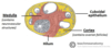
Describe the medulla of the ovary?
- This is the inner part of the ovary
- Comprised of stroma, containing the neurovascular stuctures (e.g. arteries, veins, nerves)

Describe the cortex of the ovary?
- Outer part of the ovary
- Composed of developing follicles (at various stages of development)

What kind of epithelium surrounds the ovaries?
Simple cuboidal epithelium
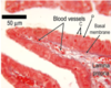
What are the four tunics of the uterine tube (from inner to outer layers)?
- Tunica mucosa (inner layer)
- Tunica submucosa
- Tunica muscularis
- Tunica serosa
What part of the female reproductive tract is this histological image

The uterine tubes
Describe the structure of the tunica mucosa of the uterine tube?
- Highly folded around the length of the tube.
- Varies from different regions- ampulla is more elaborate folded.
- The isthmus is less elaborately folded
What are the sublayers of the tunica mucosa of the uterine tube?
Epithelium
Basement membrane
Lamina propria
What is the epithelium of the uterine tubes?
Simple columnar epithelium that has two different cell types.
Peg cells (also known as secretory cells).
Ciliated cells
What is the functions of the ciliated cells in the tunica mucosa layer of the uterine tube?
Beats in one direction (the direction towards the uterus)
Has two functions:
- Aids in the movement of the ovum from the ovary to the uterus
- Protects against infection but beating pathogens away from the uterus.
Describe the tunica muscularis layer of the uterine tube?
Consists of two layers:
- Circular layer of smooth muscle (inner layer)
- Longitudinal layer of smooth muscle (outer layer).
Thick layer of smooth muscle, which contracts to help move the sperm along the uterine tube.
Thickest in the isthmus- as this is the only part prior to the ampulla- where fertilsiation occurs
What kind of movement of the tubal musculature in the uterine tube helps move the sperm along the uterine tube to the ampulla?
Peristaltic movement i.e. progressive wavelike contractions
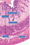
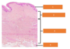
What are the 3 layers of the uterus?
- Endometrium (inner layer)
- Stratum functionalis
- Stratum basalis
- Myometrium (middle layer)
- Peritoneum (outer layer)

Name these layers of the uterus?

Compare the stratum functionalis and basalis?
Stratum functionalis
- Superficial layer
- Proliferates in response to oestrogen
- Becomes secretory in response to progesterone
- Shed during menstruation
- Blood supply is coiled arteries.
Stratum basalis:
- Deep layer
- More cellular
- Little to no change throughout the menstrual cycle
- Maintained during menstruation
- Blood supply is straight arteries.
NOTE: both contain uterine glands
The stratum functionalis becomes secretory in response to which hormone?
Progesterone
The stratum functionalis proliferates in response to which hormone?
Oestrogen
What is the blood supply to the stratum functionalis?
Coiled arteries
What is the blood supply to the stratum basalis?
Straight arteries
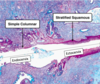
Describe the histology of the cervix region?
- The transition of epithelium occurs here.
- Two parts of the cervix:
- Endocervix
- Ectocervix
Compare the endocervix and ectocervix region?
Endocervix:
- Upper part of the cervix
- Lined by simpled columnar epithelium
- Contains mucous-secreting glands.
Ectocervix:
- Lower part of the cervix
- Lined by stratified squamous epithelium
The transition point between endocervix and ectocervix is called what?
External os
The endocervix is lined by which epithelium?
Simple columnar epithelium
The ectocervix is lined by which epithelium?
Stratified squamous epithelium
Name these regions in the cervix?

The peritoneum layer in the cervix is continous which what structure?
The peritmetrium (aka the abdominal peritoneum)
Describe the myometrium layer of the uterus?
- Middle and the thickest layer of the uterus wall.
- Composed of smooth muscle.
- The smooth muscle cells undergo hypertrophy and hyperplasia during pregnancy in preparation to expel the foetus at birth.
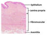
What are the four layers of the vagina, from inner to outer layer?
- Epithelium (inner layer)
- Lamina propria
- Fibromuscular layer
- Adventitia (outer layer)
Name these layers of the vagina?

What type of epithelium lines the vagina?
Stratified sqamous non-keratinised epithelium
The vagina is lubricated by mucus. From which location is this mucus coming from?
Cervical mucus lubricates the vagina.
The vagina does not contain glands
What are the 3 phases of the ovarian cycle?
The ovarian cycle represents the changes that occur in/from the ovaries.
- Follicular phase
- Ovulation
- Luteal phase
How long does the follicular stage last?
Around 14 days however varies significantly between women
Describe what happens during the follicular phase?
- At the beginning of each menstrual cycle, 15 to 20 primordial follicles are stimulated to grow under the influence of FSH
- The follicles develop until one reaches the tertiary stage.
The follicles are located at what part of the ovary?
Cortex (outside bit)
The follicles develop in stages. Name these stages?
- Primordial follicle
- Primary follicle
- Secondary follicle
- Tertiary follicle
What is the name of the follicles that are in the arrested state?
Premordial follicle
Describe the structure of the primordial follicles?
Contains a primary oocyte, which is surrounded by a single layer of squamous (flattened) follicular cells.

Which follicle does the primordial follicle develop into?
Primary follicle
The primary oocyte is arrested in which stage of development?
Arrested in meiosis I.
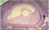
This histological image represents which stage of follicular development?
Name these parts

Primordial follicle

Describe the characteristics of the primary follicle?
- Surrounded by stratified follicular cells
- Follicular cells are now termed granulosa cells.
- Stratified: the follicular cells start to proliferate forming multiple layers i.e. multiple layers of granulosa cells.
- The oocyte secretes glycoproteins, which condense around it forming the zona pellucida.
- Moreover, at this stage the theca cells are being recruited from the surrounding stroma

This is an example of which kind of follicle?

Primary follicle
Name these parts of the primary follicle?


Describe how the zone pellucida is formed? which stage of follicular development does it form
The oocyte secretes glycoproteins, which condense around it forming the zona pellucida.
Formed during the primary follicle stage
What kind of follicles are represented by A,B and C?

A- Secondary follicle
B- Primordial follicle
C- Primary follicle
What is the structure shown in this image?

Secondary follicle
What cells are respresented by A, B and C in this image?
A- Theca interna
B- Theca externa
C- Granulosa cells
What substance is produced by the cells labelled C?

Aromatase enzymes- to convert androgens into oestrogen
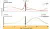
Describe the The Hypothalamic-Pituitary-Gonadal (HPG) Axis relationship during the follicular stage?
- The low level of oestrogen and progesterone inhibits the release of gonadotropin and the androgen hormones (LH and FSH) via negative feedback loops.
- Therefore, levels of FSH and LH remain low

Describe the hormonal changes that trigger ovulation?
- As the follicle matures, it releases higher levels of oestrogen. Eventually, the level of oestrogen is high enough it switches off the negative feedback and switched on the positive feedback. The positive feedback stimulates the release of GnRH, FSH and LH.
- During the negative feedback, the anterior pituitary still produced LH and FSH but did not release them. Therefore, when the feedback loop is switched to positive, there is a surge in LH
- A surge in LH triggers the rupture of the dominant follicle, releasing the secondary oocyte. It does this by stimulating enzymes that initiate the breakdown of the follicle wall.

Describe the The Hypothalamic-Pituitary-Gonadal (HPG) Axis relationship during the luteal stage?
- The lutein cells of the corpus luteum produce mainly progesterone and a small amount of oestrogen.
- The levels of oestrogen and progesterone inhibit the secretion of LH and FSH via the negative feedback loop
- Low level of oestrogen switches the feedback loop to negative
- High levels of progesterone highly inhibit the release of FSH and LH.
- Therefore, low levels of FSH and LH in this phase
The proliferative phase runs alongside which phase in the ovarian cycle?
Follicular phase

The secretory phase runs alongside which phase in the ovarian cycle?
Luteal phase

Name the uterine cycle phases the pictures correspond to?


During a normal menstrual cycle, through which three phases does the endometrium pass and on which days of the cycle do they occur?

Describe the hormonal changes that are occuring in the proliferatory/follicular stage of the mentruation cycle?
- In this stage the follicles are developing
- As they develop, the follicle produces mainly oestrogen and progesterone
- The oestrogen and progesterone, through the negative feedback loop, inhibiting the release of GnRH, LH and FSH.

Describe the hormonal changes that are occuring in the pre-ovulation stage (around day 12) of the mentruation cycle?
- Follicles developing increasing the levels of oestrogen they are producing.
- A high level of oestrogen switches the negative feedback to positive.
- Positive feedback loop= increasing the release of LH and FSH.

Describe the hormonal changes that are occuring in the luteal/secretory stage of the mentruation cycle?
- Corpus luteum produces mainly progesterone and a little of oestrogen.
- A low level of oestrogen switches the positive feedback loop to negative.
- Inhibition of the release of GnRH, LH and FSH
