Exam III - Cardiovascular System Flashcards
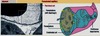
Identify ‘A’

Right ventricle
Identify ‘B’

Interventricular septum
Identify ‘C’

myocardium
Identify ‘D’

Left ventricle
Identify ‘E’

endocardium
Identify ‘F’

aorta
Identify ‘G’

right atrium
Identify ‘H’

epicardium
Identify the zone labeled ‘A’ in this H&E section of the heart

epicardium = Mesothelium, connective tissue, vessels and epithelium
Identify the zone labeled ‘B’ in this H&E section of the heart

myocardium = muscle mass
Identify the zone labeled ‘C’ in this H&E section of the heart

Endocardium=Endothelial lining of the heart chambers/surface of the valves
Purkinje fibers are found in this area
Identify ‘A’ in this H&E section of the heart

myocardium
Identify ‘B’ in this H&E section of the heart

endocardium
What are the structures circles in brown?
What is the brown pigment indicated by the blue arrow?
(this is a section of myocardium)

1 - intercalated discs
2 - lipofuscin (wear and tear pigment)
What is indicated by the arrow labeled ‘A’?

a vein
What is indicated by the arrow labeled ‘B’?

an artery
Identify the structures labeled ‘A’ and ‘B’

A - arteriole
B - nucleus of the arteriole (protrudes toward the lumen a bit)
Identify the region labeled A in this H&E section of an artery.

tunica intima
Identify the region labeled B in this H&E section of an artery.

tunica media
smooth muscle cells producing elastic, reticular, and collagenous fibers
Identify the region labeled C in this H&E section of an artery.

tunica adventitia
loose connective tissue, blood vessels, lymphatics, and nerves
Identify the region labeled ‘A’ in this H&E section of the aorta

tunica intima
Identify the region labeled ‘B’ in this H&E section of the aorta

tunica media
Identify the region labeled ‘C’ in this H&E section of the aorta

tunica adventitia
Identify the region labeled ‘A’ on this H&E section of a muscular artery

Tunica adventitia
Identify the region labeled ‘B’ on this H&E section of a muscular artery

Tunica media
Identify the region labeled ‘C’ on this H&E section of a muscular artery

tunica intima
Identify the cells indicated by the ‘A’ arrows in this longitudinal section of an arteriole

smooth muscle cells
Identify the structures indicated by the ‘B’ arrows in this longitudinal section of an arteriole

smooth muscle nuclei
Where are we likely to find fenestrated capillaries?
In tissues with substantial fluid exchange, e.g.: intestinal villi, choroid plexus, ciliary process, glomerular capillaries.
Where are we likely to find continuous capillaries?

Muscle, brain, bone, Lung, etc.
E.g.: Bloodbrain and blood-testis barriers.
Where are we likely to find discontinuous (sinusoidal) capillaries?

Hepatic and splenic sinusoids –> large molecules can exit (RBC in the spleen).
Identify ‘1’ in this TEM section of a continuous capillary.

nucleus of endothelial cell
Identify ‘2’ in this TEM section of a continuous capillary.

Pinocytotic vesicles
Identify ‘3’ in this TEM section of a continuous capillary.

tight junctions
Identify ‘4’ in this TEM section of a continuous capillary.

basement membrane/lamina
What the hell is that?! Huh?!

It’s a capillary.
Just a reminder of how majestic you’re going to look when you’re walking into 2nd semester after passing all of your exams!

That’s right… fuckin’ majestic.
What are the orange spots in this section of a renal corpuscle?

erythrocytes in glomerular capillaries
What is shown in this image?

hepatic sinusoids
Identify the structure labeled A

vein
Identify the structure labeled B

artery
Identify the structure labeled C

lymphatic vessel
no RBCs present in lymph, so it appears clear