Wk 6 GI Embryology & Anatomy/Phys Flashcards
The primitive gut tube is derived from the extraembryonic part of embryo’s yolk sac. True or false.
False
Extraembryonic yolk sac = 2ndary yolk sac –> provides nutrients to the embryo while utero-placental circulation is estsablished; later assimilited into umbilical cord.
Intraembryonic yolk sac = primitive gut
How is the gut tube formed from the 3 germ cell layers?
Endoderm: epithilial lining of digestive tube & digestive organs (liver, gallbladder, & pancreas) arise as buds
Mesoderm: through lateral folding, eventually surrounds the gut tube forming connective tissue & muscular walls.
Ectoderm: forms the neural tube that gives rise to the neural crest cells that invade the mesoderm forming neurons & glial cells intrinsic to GI tract

What are the 3 parts of the endodermal gut tube?
Foregut, midgut, & hindgut
What parts of the digestive system arises from the foregut?
Esophagus, stomach, proximal duodenum (to ampulla of Vater)
Liver/biliary apparatus
Pancreas
What artery supplies blood to the foregut?
Celiac

What parts of the digestive system arise from the midgut?
Small intestine (including duodenum distal to the bile duct)
Part of the large intestine: cecum, appendix, ascending colon & proximal transverse colon (2/3 of transverse colon)

What artery supplies blood to the midgut?
Superior mesenteric

What parts of the digestive system arise from the hindgut?
Large intestines: distal transverse colon, descending colon, sigmoid colon, rectum, superior part of anal canal
What parts of the endodermal gut tube (foregut, midgut, & hindgut) give rise to organs that are not part of the digestive system?
Foregut = Lower respiratory system & pharynx –> in the book known as a 4th section of the primitive gut, the Pharyngeal gut (includes the 2 mentioned above & the upper esophagus), the foregut is the lower esophagus down
Hindgut = epithelium of urinary bladder & most of urethra
What eventually forms into the liver, gallbladder, and pancreas?
Duodenal buds
By the 4th-6th week, what is a major early hematopoietic organ of the embryo?
Liver
When does the bone marrow take over hematopoiesis from the liver?
Approx 6 months
At birth a child’s liver is equivalent to an adult’s size. True or false.
False, at birth, the liver is 20% of the adult size –> will continue to grow for 25-30 years.
What attaches the liver to the ventral wall?
Falciform ligament

What is the respiratory diverticulum?
Lung bud
In the embryo, arising frm the foregut, the respiratory diverticulum grows from the ventral wall of the esophagus at its border with the pharyngeal gut.
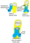
What is the esophagotracheal septum?
The separation between the respiratory diverticulum & the foregut portion that eventually becomes the esophagus

What are the 4 layers of the GI tract? (inside to outside, include the sublayers)
Mucosa, submucosa, muscularis, & serosa

Mucosa (epithelium, lamina, muscularis mucosae)
Submucosa
Muscularis (Circular muscle layer, longitudinal muscle layer)
Serosa (connective tissue layer, peritoneum)
By which week of gestation does the respiratory diverticulum separate from the foregut that eventually becomes the esophagus?
Wk 4
What are the steps forming the enteric nervous system?
1) neural crest cells enter the gut
2) crest cells proliferate
3) crest cells migrate along the gut
4) crest cells differentiate & form connections w/ their targets.
How is the GI tract innervated?
ANS - sympathetic slows it down, parasympathetic (vagus nerve) speeds it up
Also have intrinsic innervation - submucosal plexus (Meissner plexus) & myenteric plexus (Auerbach plexus)
Describe the innervation of the stomach.
Extrinsic - originate outside stomach –> parasympathetic fibers frm vagus nerve & sympathetic fibers frm the celiac plexus
Intrinsic - originate w/in stomach & respond to local stimuli –> myenteric plexus

What is peristalsis?
Coordinated sequential contraction & relaxation of the outer longitudinal & inner circular layers of muscles.
What gestation age does the gut develop normal propulsive motility/peristalsis?
30 wks
What gestational age can you swallow?
11-12 wks
What gestational age does non-nutritive sucking develop?
18-24 wks
What gestational age does coordinated esophageal peristalsis occur?
32 wks
What gestational age does nutritive sucking occur?
34-35 wks when there is rapid growth of the fetal stomach & small intestine motility
What is the percentage of the blood supplied by hepatic artery and portal vein in the liver?
hepatic artery = 25%
portal vein = 75%
What is deglutition?
the act of swallowing
What are gastric glands and gastric pits? Location.
Location: stomach’s mucosa
Gastric pits: depressions in the epithelial lining of the stomach –> a duct where gastric glands empty into.
Gastric glands: found at the bottom of gastric pits - tubular in nature –> chief cells secrete gastric juice, parietal cells secrete stomach acid.

What are parietal cells? Location & function.
Location: stomach mucosa, fundus & body
Function: cells w/in the gastric gland that secrete hydrochloric acid (gastric acid) & intrinsic factor

What are chief cells? Location & function.
Location: stomach’s mucosa, fundus & body
Function: cells w/in the gastric gland that secrete pepsinogen & gastric juices

What is pepsinogen and pepsin? Source, action, & stimulus in the GI system?
Source: chief cells in stomach
Pepsinogen: enzyme precursor to pepsin; turns into pepsin at pH of 2
Pepsin: enzyme in gastric juice that degrades food proteins into peptides
Stimulus: acetylcholine through vagal nerve stimulation during cephalic & gastric phases
Describe the development of the intestine in the fetus & the gestational week.
Wk 4: begins as a single tube
Wk 5-9: tube elongates & herniates into unbilical cord & starts to rotate. Villi are formed in jejunum
Wk 10: tube reenters the abdominal cavity the rotates -270 degrees. Microvilli & crypts of lieberkuhn appear.
Wk 13: muscularis & muscle layers well dvlped
Wk 14: Villi throughout the intestine
When is meconium present in the fetus?
Wk 16
What are the parts of the small intestine?
Duodenum, jejunum, & ilem.
Duodenum begins at pylorus & ends at the Treitz ligament where it joins the jejunum. The end of jejunum & beggining of ileum not distinguished by anatomic marker, but jejunum slightly larger lumen than the ileum.

What is the Treitz ligament?
Suspensory muscle of the duodenum - connects the duodenum to the diaphragm.
Important anatomical landmark of duodenojejunal junction

How are the 3 parts of the small intestine suspended or attached?
Duodenum: lies behind the peritoneum (retroperitoneal cavity) & attached to posterior abdominal wall.
Ileum & Jejunum: suspended in loose folds frm the posterior abdominal wall by the mesentary.

What is the peritoneum? What are the 2 types?
Serous membrane surrounding the organs of the abdomen & pelvic cavity
Visceral peritoneum - lies over organs
Parietal peritoneum - lines walls of abdominal cavity

What is the mesentery? Location.
Location: abdominal cavity
A peritoneal membrane that suspends the ileum & jejunum loosely frm the posterior abdominal wall.
Facilitates intestinal motility & supports blood vessels, nerves, & lymphatics.

What artery supplies blood to the small intestine?
Duodenum - gastroduodenal artery
Jejunum & ileum - superior mesenteric artery

What is plica? Location
Location: small intestine
Mucosal folds that slow the passage of food = more time for digestion & absorption
What are villi? Discuss location, function, & anatomy
Location: small intestine mucosa
Function: Cover the plica (mucosal folds) and secrete enzymes for digestion & absorb nutrients. At sites called tight junctions (where the columnar cells closely adhere to each other), water & electrolytes are absorbed.
Anatomy: composed of enterocytes (absorptive columnar cells) & goblet cells (mucus-secreting)
Each columnar cell has microvilli –> increases surface area for absorption.

What is the brush border? Location.
Location: small intestine
Simple columnar epithleium covered by microvilli.
Has a coating of unstirred layer of fluid important for absorbtion of substances other than water & electrolytes.

What are crypts of Lieberkuhn? Location & function.
Location: space between the bases of the villi of the small intestine.
Function: extend to the submucosal layer; where undifferentiated (stem cells) & secretary cells & Paneth cells are located.

What are Paneth cells? Location & function.
Location: at the bottom of the crypts of Lieberkuhn found in btwn villi of the small intestine.
Function: protects the small intestine - secretes antimicrobial peptide alpha-defensin 5

Where do proteins, carbohydrates, & fats go after absorption?
Protein & carbohydrates broken down to amino acids & monosaccharides –> villus capillary –> hepatic portal vein –> liver
Fat broken down to monoglycerides & long-chain fatty acids –> lacteals –> thoracic duct –> systemic circulation –> liver
Fat broken down to short-chain fatty acids & glycerol –> villus capillaries –> portal vein –> liver

What is a lacteal? Location & function
Location: within the villi of the small intestine.
Function: a lymphatic channel that absorbs & transports fat molecules.

What is the ileocecal valve/sphincter?
Location: btwn ileum & cecum of large intestine.
Controls flow of chyme & prevents reflux into the small intestine
Normally closed but peristaltic waves cause it to open, allowing small amt of chyme to pass through
Intrinsically regulated

Name the sphincters & location of the digestive system from the mouth to the anus?
Upper esophageal sphincter (Cricopharyngeal muscle) - prevents entry of air into the esophagus during respiration.
Lower esophageal sphincter (Cardiac sphincter) - prevents regurgitation frm the stomach.
Pyloric sphincter - stomach to duodenum
Ileocecal sphincter - ileum to cecum of large intestines
O’Beirne sphincter - sigmoid colon to rectum
Anal sphincter (internal/external)

What is the ampulla of Vater?
The union of the common bile duct of the liver & the pancreatic duct.
It empties into the duodenum through the sphincter of Odi
The pathway of bile secretion

Where do proteins, carbohydrates, & fats go after absorption?
Protein & carbohydrates broken down to amino acids & monosaccharides –> villus capillary –> hepatic portal vein –> liver
Fat broken down to monoglycerides & long-chain fatty acids –> lacteals –> thoracic duct –> systemic circulation –> liver
Fat broken down to short-chain fatty acids & glycerol –> villus capillaries –> portal vein –> liver
