Wk 6 GI Flashcards
Except for chewing, swallowing, and defecation, how are the movements of the digestive system (GI motility) controlled?
Autonomic nervous system (sympathetic & parasympathetic) & hormones
What are the 4 layers of the GI tract? (inside to outside, include the sublayers)
Mucosa, submucosa, muscularis, & serosa
Mucosa (epithelium, lamina, muscularis mucosae)
Submucosa
Muscularis (Circular muscle layer, longitudinal muscle layer)
Serosa (connective tissue layer, peritoneum)

What are the 3 enteric nerve plexuses and their locations?
Submucosal plexus, myenteric plexus, & subserosal plexus
Submucosal plexus (Meissner plexus) - in the muscularis mucosae
Myenteric plexus (Auerbach plexus) - in btwn circular & longitudinal muscle layers
Subserosal plexus - beneath the serosa

How many permanent adult teeth in the mouth?
32
What are the 3 salivary glands?
Parotid, submandibular, & sublingual

How much saliva is secreted per day?
1 L
What is saliva made up of?
Mucus
Electrolytes (sodium, bicarbonate, chloride, potassium)
Ptyalin (salivary alpha-amylase)
What is ptyalin?
Salivary amylase - an enzyme that digests carbohydrates in mouth and stomach
What stimulates and inihibits salivary glands?
ANS controls salivation.
Cholinergic parasympathetic fibers & beta-adrenergic sympathetic fibers stimulate
Atropine (anticholinergic agent) inhibits salivation & makes it dry
Are salivary glands regulated by hormones?
No
How does saliva prevent tooth decay?
Bicarbonate has pH of 7.4 = neutralizes bacterial acids
IgA (immunoglobulin A) prevents infection (also found in saliva)
Exogenous fluoride (fluoride in drinking water) absorbed & excreted in saliva
How does swallowed food move from the mouth to the stomach?
Esophageal peristalsis
What is peristalsis?
Coordinated sequential contraction & relaxation of the outer longitudinal & inner circular layers of muscles.
Describe the muscles and nerves in the esophagus.
Upper = striated muscle innervated by motor neurons
Middle = mix of striated & smooth muscle
Lower = smooth muscle innervated by preganglionic cholinergic fibers frm the vagus nerve

How is peristalsis in the esophagus stimulated?
As food passes & causes stretching of the walls, afferent fibers sense changes in the wall tension.
The greater the tension, the greater the esophageal contraction.
Name & descibe the function of the esophageal sphincters.
Upper esophageal sphincter (cricopharyngeal muscle) & lower esophageal sphincter (cardiac sphincter)
Upper esophageal sphincter (Cricopharyngeal muscle) - prevents entry of air into the esophagus during respiration.
Lower esophageal sphincter (Cardiac sphincter) - prevents regurgitation frm the stomach.
How is swallowing controlled?
Swallowing center in the brainstem
Name & describe the 2 phases of swallowing.
Oropharyngeal & esophageal
Oropharyngeal (voluntary) phase - food bolus forced posteriorly by the tongue toward the pharynx. Superior constrictor muscle of the pharynx contracts, preventing movement of food into the nasopharynx. As respiration is inhibited, the epiglottis slides downard to prevent the bolus frm entering larynx & trachea.
Esophageal (involuntary) phase - food bolus enters the esophagus. Primary peristalsis occurs. If bolus of food b/comes stuck, 2ndary peristalsis occurs. Peristalsis = wave of relaxation (allows food to pass & reduces resistance), then wave of contraction (pushes food along).
What is partially digested food called?
Chyme
Describe the anatomic structure of the stomach.
Boundaries of the stomach: lower esophageal sphincter (cardiac sphincter), greater & lesser curvatures, pyloric sphincter
Functional areas of the stomach: fundus (upper portion), body (middle portion), & antrum (lower portion)

Name the 3 smooth muscle layers of the stomach (inside to outside)
Oblique layer
Circular layer
Longitudinal layer

Which parts of the stomach does the muscle layers become progressively thicker?
Body & antrum b/c ths is where food is mixed, churned, & pushed out to tthe duodenum

What artery supplies blood to the stomach?
Celiac artery
Describe the veins of the stomach
Splenic vein drains R side of stomach
Gastric vein drains L side of stomach

Describe the innervation of the stomach.
Extrinsic - originate outside stomach & controlled by vagus nerve & branches of the celiac plexus –> parasympathetic fibers frm vagus nerve & sympathetic fibers frm the celiac plexus
Intrinsic - originate w/in stomach & respond to local stimuli –> myenteric plexus

Can the stomach absorb substances? Give ex. of what the stomach absorbs.
Few substances can be absorbed in stomach - mucosa impermeable to water.
Can absorb alcohol & aspirin.
What action causes the fundus to relax making it more ceptive to receive a bolus of food?
Swallowing
What 2 intestinal hormones increase gastric motility/contraction? How?
Gastrin & motilin - make the threshold potential of muscle fibers less negative
Does the vagus nerve increase or inhibit gastric motility?
Increase
What inhibits gastric motility by making the threshold potential more negative?
Sympathetic activity & secretin (intestinal hormone)
What is gastrin? Include source, action, stimulus for secretion
Source: G cell of stomach’s mucosa
Action: a hormone stimulates parietal cells to secrete hydrochloric acid (gastric acid) and chief cells to secrete pepsinogen; promotes gastric motility & regulates gastroileal reflex
Stimulus for secretion: partially digested proteins in stomach
What is motilin? Include source, action, & stimulus for secretion.
Source: small intestine
Action: a hormone that increases GI motility
Stimulus for secretion: acid or fat in the duodenum
When does the pyloric sphincter open during digestion?
Trick question: pylorus is always open about 2mm
It opens wider during antral contraction - normally no regurgitation frm duodenum to stomach.
What increases the rate of gastric emptying?
Larger volumes of food –> increases gastric pressure, peristalsis, & rate of emptying
What decreases gastric emptying?
Solids, fats, & nonisotonic solutions
What hormone inhibits gastric motility & decreases gastric emptying?
Cholecystokinin
What is cholecystokinin? Include source, action, & stimulus for secretion.
Source: small intestine
Function: stimulates gallbladder to eject bile & pancrease to secrete alkaline fluid; decr. gastric motility & delays gastric emptying so that fats are not emptied into the duodenum at a rate that exceeds the rate of bile & enzyme secretion. Inhibits gastrin; constricts pyloric sphincter; regulates gastroileal reflex
Stimulus for secretion: presence of chyme (acid, partially digested proteins, fats) in the duodenum
How is the stomach’s peristaltic activity affected by blood glucose levels?
Low blood glucose levels stimulate vagus nerve & gastric smooth muscles to increase peristalsis, but not emptying –> stimulates the sensation of hunger pains.
What is the function of mucus in the stomach? Stimulus?
Protective barrier against acid & proteolytic enzymes
Stimulus: prostaglandins & nitric oxide –> also stimulate bicarb & inhibits secretion of acid, further protecting the stomach.
What are gastric glands and gastric pits? Location.
Location: stomach’s mucosa
Gastric pits: depressions in the epithelial lining of the stomach –> a duct where gastric glands empty into.
Gastric glands: found at the bottom of gastric pits - tubular in nature –> chief cells secrete gastric juice, parietal cells secrete stomach acid.

What are parietal cells? Location & function.
Location: stomach mucosa, fundus & body
Function: cells w/in the gastric gland that secrete hydrochloric acid (gastric acid) & intrinsic factor

What are chief cells? Location & function.
Location: stomach’s mucosa, fundus & body
Function: cells w/in the gastric gland that secrete pepsinogen & gastric juices

What is pepsinogen and pepsin? Source, action, & stimulus in the GI system?
Source: chief cells in stomach
Pepsinogen: enzyme precursor to pepsin; turns into pepsin at pH of 2
Pepsin: enzyme in gastric juice that degrades food proteins into peptides
Stimulus: acetylcholine through vagal nerve stimulation during cephalic & gastric phases
What are G cells? Location & function
Location: stomach’s mucosa in the gastric pit, part of gastric gland; toward the antrum
Function: Secretes gastrin

What are enterochromaffin-like cells? Location & function.
Location: stomach’s mucosa in the gastric pit, part of gastric gland
Function: secrete histamine

What are D cells? Location & function.
Location: stomach’s mucosa in in the gastric pit, part of gastric gland
Function: secrete somatostatin

What are the parts of the small intestine?
Duodenum, jejunum, & ilem.
Duodenum begins at pylorus & ends at the Treitz ligament where it joins the jejunum. The end of jejunum & beggining of ileum not distinguished by anatomic marker, but jejunum slightly larger lumen than the ileum.

What is the Treitz ligament?
Suspensory muscle of the duodenum - connects the duodenum to the diaphragm.
Important anatomical landmark of duodenojejunal junction

What is the ileocecal valve/sphincter?
Location: btwn ileum & cecum of large intestine.
Controls flow of chyme & prevents reflux into the small intestine
Normally closed but peristaltic waves cause it to open, allowing small amt of chyme to pass through
Intrinsically regulated

What is the peritoneum? What are the 2 types?
Serous membrane surrounding the organs of the abdomen & pelvic cavity
Visceral peritoneum - lies over organs
Parietal peritoneum - lines walls of abdominal cavity

How are the 3 parts of the small intestine suspended or attached?
Duodenum: lies behind the peritoneum (retroperitoneal cavity) & attached to posterior abdominal wall.
Ileum & Jejunum: suspended in loose folds frm the posterior abdominal wall by the mesentary.

What is the mesentery? Location.
Location: abdominal cavity
A peritoneal membrane that suspends the ileum & jejunum loosely frm the posterior abdominal wall.
Facilitates intestinal motility & supports blood vessels, nerves, & lymphatics.

What artery supplies blood to the small intestine?
Duodenum - gastroduodenal artery
Jejunum & ileum - superior mesenteric artery

Where does the superior meseneteric vein empty into?
The superior mesenteric vein & splenic vein empties into the portal circulation to the liver.

Describe the muscles of the small intestine
Smooth muscles of small intestine = circular inner layer & longitudinal outer layer

What is plica? Location
Location: small intestine
Mucosal folds that slow the passage of food = more time for digestion & absorption

What are villi? Discuss location, function, & anatomy
Location: small intestine mucosa
Function: Cover the plica (mucosal folds) and secrete enzymes for digestion & absorb nutrients. At sites called tight junctions (where the columnar cells closely adhere to each other), water & electrolytes are absorbed.
Anatomy: composed of enterocytes (absorptive columnar cells) & goblet cells (mucus-secreting)
Each columnar cell has microvilli –> increases surface area for absorption.

What is the brush border? Location.
Location: small intestine
Simple columnar epithleium covered by microvilli.
Has a coating of unstirred layer of fluid important for absorbtion of substances other than water & electrolytes.

What is a lacteal? Location & function
Location: within the villi of the small intestine.
Function: a lymphatic channel that absorbs & transports fat molecules.

What are crypts of Lieberkuhn? Location & function.
Location: space between the bases of the villi of the small intestine.
Function: extend to the submucosal layer; where undifferentiated (stem cells) & secretary cells & Paneth cells are located.

What are Paneth cells? Location & function.
Location: at the bottom of the crypts of Lieberkuhn found in btwn villi of the small intestine.
Function: produce defensins & other abx peptides & proteins.

Where do proteins, carbohydrates, & fats go after absorption?
Protein & carbohydrates broken down to amino acids & monosaccharides –> villus capillary –> hepatic portal vein –> liver
Fat broken down to monoglycerides & long-chain fatty acids –> lacteals –> thoracic duct –> systemic circulation –> liver
Fat broken down to short-chain fatty acids & glycerol –> villus capillaries –> portal vein –> liver

How is water transported in the small intestine?
Transported through the tight junction & intercellular spaces vs across cell membranes
Diffuses passively according to hydrostatic pressure & in relation to osmotic gradients of electrolytes

Describe how protein, fat, and carbohydrates are absorbed/transported across the epithelial cell of the villus.
Protein broken down to amino acids –> active transport or 2ndary active transport w/ Na –> villus capillaries –> portal vein –> liver
Protein broken down to dipeptides & tripeptides –> 2ndary transport w/ H+ –> –> villus capillaries –> portal vein –> liver
Carb broken down to glucose & galactose –> 2ndary active transport with Na –> villus capillaries –> portal vein –> liver
Carb broken down to fructose –> facilitated diffusion –> villus capillaries –> portal vein –> liver
Fat emulsified to short-chain fatty acids & glycerol –> simple diffusion –> villus capillaries –> portal vein –> liver
Fat emulsified into micelle (makes it water-soluble) which turns into long-chain fatty acids & monoglycerides –> simple diffusion –> b/comes a triglyceride in the cell which turns into a chylomicron –> lacteal of a villus –> thoracic duct –> eventually reach the liver through systemic circulation

How is sodium transported across the epithelial cell of the small intestine?
Diffuses through tight junctions –> proximal part of intestine more permeable than distal part
Active transport –> transported into intestinal cells in exchange for hydrogen at brush border. To maintain electroneutrality in the ileum, chloride actively enters cell in exchange for bicarbonate.
Secondary active transport –> Sodium pump at basolateral membrane –> glucose transport enhances sodium absorption.

What are the enzymes that break down carbohydrates? Discuss source & site of action.
Salivary amylase (breaks down starch to dextrins & oligosaccharides) –> source: mouth, site of action: mouth
Pancreatic amylase (breaks down dextrins & oligosaccharides to lactose, maltose, & sucrose) –> source: pancreas, site of action: small intestine
Lactase, Maltase, & Sucrase (breaks down lactose, maltose, & sucrose to galactose, glucose, & fructose) –> source: brush-border of small intestine, site of action: small intestine

What agents & enzymes break down fats? Discuss site of action & source
Bile acids (emulsifies fat) –> source: liver, site of action: small intestine
Pancreatic lipases (breaks triglycerides into monoglycerides & long-chain fatty acids OR glycerol & short-chain fatty acids)–> source: pancreas, site of action: small intestine

How does the small intestine absorb iron?
Iron bound to intestinal transferrin in small intestine –> absorbed in epithelial cell & bound to ferritin –> transported to blood by plasma transferrin

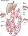
What are the parts of the large intestine?
Cecum, appendix, colon, rectum, & anal canal

What is the cecum? Location.
Location: part of the large intestine
A pouch that receives chyme frm the ileum & attached to it is the vermiform appendix

What are the 4 parts of the colon?
Ascending colon, transverse colon, descending colon, & sigmoid colon.

What is the internal & external anal sphincter? Location & innervation.
Location: end of anus, part of the large intestine
Internal anal sphincter: involuntarilty regulates fecal passage; part of the inner surface of the anal canal; composed of circular muscle layers –> innervated by parasympathetic fibers
External anal sphincter: –> voluntarily regulates fecal passage; part of the encircling outside wall of the anal canal & canal opening; composed of striated muscle –> innervated by pudendal nerve (carries motor & sensory fibers)

Name the sphincters & location of the digestive system from the mouth to the anus?
Upper esophageal sphincter (Cricopharyngeal muscle) - prevents entry of air into the esophagus during respiration.
Lower esophageal sphincter (Cardiac sphincter) - prevents regurgitation frm the stomach.
Pyloric sphincter - stomach to duodenum
Ileocecal sphincter - ileum to cecum of large intestines
O’Beirne sphincter - sigmoid colon to rectum
Anal sphincter (internal/external)

What are teniae coli? Location
Source: cecum & colon
3 longitudinal bands in the longitudinal muscle layer that are shorter than the colon, giving the colon it’s “gathered appearance”

What are haustra (singular haustrum)? Location
Location: cecum & colon
Outpouchings of the circular muscles of the colon

Describe the mucosa of the colon.
There are rugae (folds) btwn the haustra.
Lieberkuhn crypts, but no villi
Mucosa made up of: columnar epithelial cells that absorb fluids & electrolytes & goblet cells that secrete mucus to lubricate the mucosa
