Hip Arthroplasty (Complete) Flashcards
What are the approaches to the hip?
[Can J Surg. 2015 Apr;58(2):128-39.]
- Anterior approach (Smith-Peterson)
- Superficial plane – TFL and sartorius
- Deep plane – rectus femoris and gluteus medius
- Incision – 2cm lateral to ASIS, ~10cm
- Structures at risk:
- LFCN – take it medially
- Ascending branch of the lateral femoral circumflex artery
- Potential advantages:
- Reduced blood loss
- Earlier functional recovery
- Low dislocation rates
- Shorter stays in hospital
- Anterolateral approach (Watson-Jones)
* Plane – TFL and gluteus medius - Direct lateral approach (Hardinge)
- Plane – split of gluteus medius and vastus lateralis
- Structures at risk:
- Superior gluteal nerve (~5cm proximal to GT)
- Femoral nerve
- Posterior approach (Moore or Southern [Kocher-Langenbeck for trauma])
- Plane – split of gluteus maximus
- Incision – starts 5cm distal to tip of GT centered on femoral shaft curving 6cm proximal to GT towards PSIS
- Structures at risk
- Sciatic nerve
What is the Dorr classification of femoral bone quality?
- Determined by the canal/calcar isthmus ratio
* Endosteal width 10cm below LT / endosteal width at mid-LT - Type A
- CC ratio <0.5
- Champagne flute
- Indicates thick medial and lateral cortices and a large posterior cortex
- Femoral prosthesis suggested = press fit
- Type B
- CC ratio 0.5-0.75
- Indicates bone loss at the medial and posterior cortices
- Femoral prosthesis suggested = press fit or cemented
- Type C
- CC ratio >0.75
- Stovepipe
- Indicates a stovepipe appearance due to complete loss of both the medial and the posterior cortex and a widened intramedullary diameter
- Femoral prosthesis suggested = cemented

What is the most common indication for revision THA?
[Bone Joint J 2016;98-B(1 Suppl A):44–9]
Instability
What is the definition of early and late THA dislocators?
[Bone Joint J 2016;98-B(1 Suppl A):44–9][The Journal of Arthroplasty 33 (2018) 1316e1324]
- Early dislocator
- Dislocation within 2 years of surgery
- One study showed 59% of dislocations in first 3 months, 77% within first year
- Late dislocator
* Dislocation beyond 2 years of surgery
How do the etiologies of THA instability differ in early vs. late dislocations?
[Intl. Orth. (SICOT) (2017) 41:661–668]
- Early
* Most commonly due to malposition - Late
- Eccentric liner wear
- Implant loosening (subsidence/migration or rotation of the cup)
- Abductor muscle damage
- Greater trochanter disruption (due to osteolysis)
What are the types of THA instability described by Wera and Paprosky (Rush University Group)?
[Bone Joint J 2016;98-B(1 Suppl A):44–9]
- Type I - acetabular component malposition
- Type II - femoral component malposition
- Type III - abductor deficiency
- Type IV - soft tissue/bony impingement
- Type V - eccentric polyethylene wear
- Type VI - unknown etiology
What are the patient risk factors for postoperative THA instability?
[The Journal of Arthroplasty 33 (2018) 1316e1324]
- Age
* Bimodal distribution of greatest risk <50 and ≥70 - Obesity
- Central, spinal and neuromuscular disorders
* Eg. CP, polio, spinal injury, dementia - Spinopelvic pathology
- Poor spinopelvic mobility
- Lumbosacral fusion
- Ankylosing spondylitis
5. THA for osteonecrosis - Tend to have reduced neck shaft angle and increased femoral version
***NOTE: sex is not a risk factor
What are THA dislocation risk factors?
[JAAOS 2004;12:314-321]
- Patient factors:
- Female (2x more common than men)
- Neuromuscular and cognitive disorders
- CP, muscular dystrophy, parkinsons, psychosis, dementia, alcoholism
- THA for management of fracture
- History of surgery on the same hip (for any indication)
- Patient non-compliance with activity restrictions
- Surgical factors:
- Surgeon inexperience increases risk
- Surgical approach
- Posterior approach (increases risk of posterior dislocation)
- Soft-tissue tension
- Capsule repair reduces risk of dislocation
- Restoration of abductor tension reduces risk of dislocation
- Restore femoral offset (distance from the hip head centre to the tip of the GT or a line down the centre of the femoral canal)
- Restore femoral neck length (distance from centre of the femoral head to the base of the femoral neck – top of the LT is the usual reference)
- GT non-union or abductor avulsion increases risk of dislocation
- Component positioning
- Cup malposition
- Normal position:
- Inclination (40+/-10o)
- Anteversion (20+/-5o)
- AKA Lewinnek’s safe zone [Intl. Orth. (SICOT) (2017) 41:661–668]
- ***NOTE: this safe zone has recently been questioned and it may be more important to match to patient anatomy
- Retroversion = risk of posterior dislocation
- Excessive anteversion = risk of anterior dislocation
- Vertical cup = risk of posterior-superior dislocation
- Horizontal cup = risk of inferior dislocation
- Normal position:
- Femoral stem malposition
- Normal position = anteversion (10-15o)
- Retroversion = risk of posterior dislocation
- Excessive anteversion = risk of anterior dislocation
- ***Note: anteversion is additive between the cup and stem
- Excessive anteversion of one component may be acceptable but the additive effect if both are excessively anteverted = increased risk of dislocation
- Cup malposition
- Impingement
- Impingement of the femoral neck on a nonarticular surface can lever out of socket resulting in dislocation
- Bone = osteophyte
- Implant = neck and cup (‘intraprosthetic’)
- Soft tissue = scar
- In severe DDH it can occur between the GT and the ischium [Intl. Orth. (SICOT) (2017) 41:661–668]
- Impingement can be reduced by:
- Increasing head to neck ratio (head diameter/neck diameter)
- Increase femoral head size (also increases ‘jump distance’)
- Avoid skirts (plus size heads)
- Clearing acetabular osteophytes, extruded cement, HO
- Increasing femoral offset
- Increasing head to neck ratio (head diameter/neck diameter)
- Impingement of the femoral neck on a nonarticular surface can lever out of socket resulting in dislocation
What is the workup for THA instability?
[The Journal of Arthroplasty 33 (2018) 1325e1327]
- History
- Mechanism of instability
- Traumatic vs. positional
- Hip in flexed position = posterior dislocation (eg. getting up from chair)
- Hip in extended and ER position = anterior dislocation (eg. standing and turning)
- Antecedent pain
- Suggestive of infection, aseptic loosening, adverse local tissue reaction
- Early vs. late dislocation
2. OR notes - Surgical approach
- Components used
- Acetabulum, liner, femoral components
- Complications
- Examination
- Leg position
- Flexed, adducted, IR = posterior dislocation
- Extended, ER = anterior dislocation
- Abductor strength
- Trendelenburg sign/gait
- LLD
- Apprehension sign – indicates position at which hip becomes unstable
- Labs
* CBC, ESR, CRP - Radiographs
- AP
- Cup inclination
- Eccentric poly wear
- Head-neck ratio
- Femoral offset
- Centre of rotation
- Height and integrity of GT
- Impingement signs
- 1.Osteophytes, neck and liner, GT
- Cross-table lateral
- Cup anteversion
- Angle formed by the face of the acetabular component compared to a line perpendicular to the horizontal
6. CT scan
- Angle formed by the face of the acetabular component compared to a line perpendicular to the horizontal
- Cup anteversion
- Gold standard for assessment of acetabular and femoral component position
- Acetabular component = axial CT scan
- Angle formed by the face of the acetabular component relative to the perpendicular from a line drawn across both ischial spines
- Femoral component = axial CT scan
- Angle formed by the line parallel to femoral component through its widest metaphyseal portion relative to the ipsilateral femoral posterior condyle reference line at the distal femur
- MRI
- Angle formed by the line parallel to femoral component through its widest metaphyseal portion relative to the ipsilateral femoral posterior condyle reference line at the distal femur
- MARS – metal artifact reduction sequence
- Indicated in the setting of adverse local tissue reaction due to metal hypersensitivity to assess abductor integrity
What is the treatment of early THA instability?
Closed reduction
What is the treatment of late THA instability?
[Intl. Orth. (SICOT) (2017) 41:661–668] [Bone Joint J 2016;98-B(1 Suppl A):44–9]
- Type I – acetabular component malposition
- Revision of acetabular component
- Note – do not use elevated/lip liners in isolation to treat Type I as it increases risk of impingement, breakage and wear
- Type II – femoral component malposition
* Revision of femoral component - Type III – abductor deficiency
- Constrained liner
- Abductor repair and advancement
- Type IV – impingement
- Excise offending osteophyte, soft tissue, HO, cement
- Increase head-neck ratio
- Increase femoral head size or consider dual mobility
- Increase femoral offset
- Lateralized liner, heads with longer necks (avoid skirts), extended offset stems
- Type V – eccentric poly wear
* Modular head and liner exchange (increase femoral head size if possible) - Type VI – unknown etiology
* Constrained liner
***NOTE: in all cases consider increasing head size or dual mobility component
What are the disadvantages of constrained liners?
[Intl. Orth. (SICOT) (2017) 41:661–668]
- Acetabular and femoral component loosening
- Decreased ROM
- Increased poly wear
- Dissociation of the head from the liner (requires revision surgery)
- Liner breakage
What are the indications for constrained devices in THA?
[JAAOS 2018;26:479-488]
- Recurrent instability (three or more dislocations)
- Previous failed attempts at surgical stabilization
- Lack of identifiable causes of instability that could be corrected
- Central nervous system disorders
- Previous failure of a constrained device
- Prophylaxis in revision procedures in patients with inadequate posterior soft tissues and/or abductors
- Alcohol abuse
- Multidirectional instability intraoperatively
- Substantial abductor musculature deficiency
- Presence of an internal fixation device
- Extensive femoral or acetabular bone loss
- Inability to repair the greater trochanter
- Revision after arthrodesis or resection arthroplasty
- Revision after periprosthetic fractures
- Cognitive impairment preventing adherence to hip precautions
- Prophylaxis in the revision of metal-on-metal hip arthroplasty in patients with substantial soft-tissue defects after débridement of tissue granulomas or necrotic tissues
When considering constrained devices for THA what are the options?
[JAAOS 2018;26:479-488]
- Constrained liner
- Constrained tripolar (dual-mobility)
- Nonconstrained tripolar (dual-mobility)
What are the complications associated with dual mobility components?
- Increased poly volumetric wear
- Osteolysis
- Intraprosthetic dislocation
What is the intraoperative evaluation of combined anteversion in THA?
- With the leg positioned neutrally or with slight flexion of the hip; the leg is then internally rotated until the femoral head is symmetrically seated in the acetabular component. The degree of internal rotation represents the combined anteversion
- The safe zone for combined anteversion is 25-50° (goal of 35)
What imaging is required when managing a THA periprosthetic fracture?
[JAAOS 2009;17:677-688]
- AP pelvis
- AP and cross-table lateral of involved hip
- Full length femur views
What is the Vancouver classification of periprosthetic femur fractures?
[JAAOS 2009;17:677-688] [JAAOS 2014;22:482-490]
Classification is based on fracture location (A, B, C), stem stability, bone stock
- Type A (trochanter region)
- Vancouver AG – fracture of the GT
- Stable when minimally displaced (held by digastric tendons of vastus lateralis and glut med.)
- Usually related to wear-osteolysis of the GT
- Vancouver AL – fracture of the LT
- Vancouver AG – fracture of the GT
- Type B (diaphysis including and just distal to the tip)
- Vancouver B1 – stable implant
- Vancouver B2 – loose implant
- Vancouver B3 – loose implant + inadequate bone stock
- Type C (diaphysis well distal to the tip of the implant)
- Vancouver C – well distal to the implant + implant is stable

How do you assess for implant stability in setting of periprosthetic fracture?
[JAAOS 2009;17:677-688] [JAAOS 2014;22:482-490]
Pre-op:
- Clinical features that may suggest a loose femoral stem prior to the fracture:
- Startup pain (initiation of ambulation on rising from chair)
- Groin or thigh pain
- Pain with non-WB range of motion
- Progressive limb shortening
- Symptoms or signs of infection
- Radiographic features:
- Comparison to previous xrays
- Subsidence
- Radiolucency (cement-bone or cement-implant interface)
- Cement mantle fracture
- ***Note: cement mantle fracture alone is not indicative of a loose implant
- Bone pedestal
Intra-op:
- If distal stem is exposed attempt generating longitudinal shear (grasp femur with pointed reduction forceps and stem with kocher)
- If distal stem is not exposed then arthrotomy and hip dislocation to assess stability
What is the management of Vancouver B1 periprosthetic fractures?
[JAAOS 2009;17:677-688]
ORIF
- Locking plate
- Fixation around the stem requires screws anterior/posterior to the stem or cables
- Plate length – generally from vastus ridge (fixation in GT) to 2 cortical diameters distal to the tip of the stem or fracture site (3-5 bicortical screws distal to #)
- Minimally invasive percutaneous plating preferred when possible

What is the management of Vancouver B2 periprosthetic fractures?
[JAAOS 2014;22:482-490]
Femoral component revision
- Remove implant, cement and biomembrane
- Long-stemmed, diaphyseal fitting stem
- Monoblock, extensively porous-coated, non-cemented stem (>4-6cm of diaphyseal fixation)
- Modular, tapered stems (<4-6cm of diaphyseal fixation)
- Reconstruction of the femoral shaft
- If little comminution do prior to insertion of the stem
- If comminution exists insert the stem then reduce the fragments around the stem

What is the management of Vancouver B3 periprosthetic fractures?
[JAAOS 2014;22:482-490]
Femoral component revision
- Tumor prosthesis
- Modular tapered stem (requires <4-6cm of interference fit)
- Splines maintain rotational stability
- Cortical strut allograft
- Allograft-prosthesis composite
- Impact bone grafting

What are the indications for femoral revision in THA?
[J Am Acad Orthop Surg 2013;21: 601-612]
- Aseptic loosening
- Infection
- Osteolysis
- Periprosthetic fracture
- Component malposition
- Catastrophic implant failure
What is the Paprosky classification for femoral bone loss?
[J Am Acad Orthop Surg 2013;21: 601-612]
- Type I - minimal proximal metaphyseal bone loss
- Type II - moderate to severe proximal metaphyseal bone loss
- Type IIIA - severe proximal metadiaphyseal bone loss with ≥4cm of isthmus/diaphysis intact
- Type IIIB - severe proximal metadiaphyseal bone loss with <4cm of isthmus/diaphysis intact
- Type IV - severe metadiaphyseal bone loss with no intact isthmus/diaphysis

What are the reconstruction options for each Paprosky type of femoral bone loss?
- Type I - uncemented proximal fitting stem
- Type II - extensively porous-coated diaphyseal fitting stem
- Type IIIA - extensively porous-coated diaphyseal fitting stem
- Type IIIB - modular tapered stem
- Type IV - impaction grafting plus cemented stem OR APC stem
In the setting of loose femoral components (in THA) what is the observed proximal femoral remodeling?
[JAAOS 2013;21: 601-612]
Varus and retroversion
What is the most common approach used for femoral stem revision in THA?
[JAAOS 2013;21: 601-612]
Posterolateral
What is the indication for an ETO when revising the femurin THA?
[JAAOS 2013;21: 601-612][Instr. Course Lec. 2004]
- Significant varus remodeling
- Well-fixed uncemented stem
- Long column of cement below the stem
- Abductor tension adjustment
- Exposure of complicated acetabular reconstructions
When is an ETO indicated prior to hip dislocation (in revision THA)?
When dislocation is difficult and risks iatrogenic fracture
- HO, protrusion, ankylosis, stiffness
What is the surgical technique for an ETO?
- Posterolateral approach
* Vastus lateralis reflected anteriorly after release from linea aspera
* Osteotomy is made just lateral to linea aspera (1/3 diameter of the femoral shaft)- The length is minimum 12cm from tip of GT usually to the tip of the stem
- The distal osteotomy should not be transverse rather tapered
- The osteotomy is completed through the medial cortex
- The abductors and vastus lateralis remain attached the fragment and can be mobilized anteriorly
- Direct lateral approach [CORR 2003; 417: 210]
* As above except the anterolateral osteotomy is made first followed by the posterior osteotomy then the fragment is mobilized posterior
How is the ETO reapproximated and fixed?
- Distal Luque wire or cable to prevent propagation of osteotomy prior to implant insertion
- Multiple Luque wires or cables for final fixation
- Strut allograft if bone loss
What are the complications of trochanteric osteotomy (in revision THA)?
- Nonunion
- Migration
- Iatrogenic fracture
- Fracture of the osteotomy fragment
What instruments should be available to facilitate femoral stem removal in revision THA?
[JAAOS 2013;21: 601-612]
- Manufacturer-specific explant tools
- Flexible osteotomes
- Trephines
- High-speed burrs (eg, pencil tip, carbide tip, metal cutting wheel)
- Ultrasonic cement removal instruments
- Universal extraction tools that allow attachment to the stem or taper.
What are indications for acetabular revision in THA?
[J Am Acad Orthop Surg 2013;21]
- Aseptic loosening
- Hip instability
- Periprosthetic osteolysis
- Periprosthetic infection
What is the Paprosky Classification of acetabular bone loss in THA (based on location or migration of the hip centre of rotation, degree of teardrop destruction, amount of ischial osteolysis, integrity of Kohler line)?[J Am Acad Orthop Surg 2013;21] 128-139
Type I - minimal bone loss
Type IIA - superomedial bone loss (intact rim)
- <3cm of superior hip migration
Type IIB - superolateral bone loss (superior rim not intact)
- <3cm of superior hip migration
Type IIC - medial wall bone loss
- Moderate teardrop osteolysis and disruption of Kohler’s line
Type IIIA - “up and out”
- 30-60% superolateral rim loss (10-2 o’clock)
- >3cm superolateral hip migration
- Moderate teardrop and ischial osteolysis
- Kohler’s line intact
Type IIIB - “up and in”
- >60% acetabular bone loss (9-5 o’clock)
- Superomedial hip migration >3cm
- Severe ischial and teardrop osteolysis
- Complete disruption of Kohler’s line
- May have pelvic discontinuity
***NOTE:
- Kohler’s Line —integrity of medial wall and superior anterior column
- Acetabular Tear Drop — integrity of medial wall and inferior portion of anterior and posterior column
- Ischial Lysis —integrity of posterior wall and posterior column
- Vertical Migration —integrity of superior dome

What is the Gross Classification System of acetabular bone loss?
[J Bone Joint Surg Am. 2016;98:233-42]
- Type I - no substantial loss of bone stock
- Type II - contained loss of bone stock
- Type III - minor column defect; uncontained loss of bone stock involving <50% of acetabulum
- Type IV - major column defect; uncontained loss of bone stock involving >50% of acetabulum
- Type V - pelvic discontinuity with uncontained loss of bone stock
What is the AAOS Classification System of acetabular bone loss?
[J Bone Joint Surg Am. 2016;98:233-42]
Type I - Segmental
- Loss of part of the acetabular rim or medial wall
Type II - Cavitary
- Volumetric loss in the osseous substance of the acetabular cavity
Type III - combined deficiency
- Combination of segmental and cavitary
Type IV - pelvic discontinuity
- Complete separation between the superior and inferior aspects of the acetabulum
Type V - arthrodesis

What is the treatment of acetabular defects based on Paprosky classification?
[J Am Acad Orthop Surg 2013;21]
Type I-IIB
- Noncemented, porous-coated hemispheric implant with the use of adjunctive screw fixation
Type IIC
- As above PLUS bone grafting of the medial defect
Type IIIA
- Noncemented, porous hemispheric implant, with supplemental porous metal augments or structural graft
Type IIIB
- Noncemented acetabular device must be used in conjunction with structural allograft, a reconstruction cage, modular porous metal augments, or a combination of these options.
- May be associated with pelvic discontinuity
What is the treatment of acetabular defects based on the Gross classification?
[J Bone Joint Surg Am. 2016;98:233-42]
Type I
- Uncemented or cemented hemispherical acetabular component
Type II
- Uncemented hemispherical acetabular component and morselized bone-grafting
Type III
- Uncemented hemispherical acetabular component and minor column graft or metal augment
Type IV
- Major column graft protected by a reconstruction cage or metal augment protected by a reconstruction cage
Type V
- Cup-cage reconstruction with major column graft or metal augment
***NOTE – major column graft = allograft acetabular bone is used as a graft and shaped to fit the defect
What is the preferred approach for acetabular revision surgery?
[J Am Acad Orthop Surg 2013;21]
Posterolateral
What instrumentation is need for removal of a well fixed acetabular component?
[J Am Acad Orthop Surg 2013;21]
Explant device
- Allows for safe, quick and bone conserving removal
What are the treatment options available when reconstructing the acetabulum in revision THA?
[J Am Acad Orthop Surg 2013;21]
- Uncemented hemispheric cup +/- screw augmentation +/- bone grafting contained defects
- Uncemented hemispheric cup + structural autograft or metal augment
- Structural allograft or metal augment protected by cage + cemented liner
- Cup-cage construct +/- metal augment or structural graft
- Oblong cup
- Custom triflange
What is the definition of an antiprotrusio cage, what is the indication for same?
[J Bone Joint Surg Am. 2016;98:233-42]
- Cage that extends from ilium to ischium
* Note – more rigid than cage used in the cup cage construct - Indication – significant bone loss where uncemented hemispherical cup can not achieve stability even with use of metal augments, protection of structural allograft or cage when addressing segmental defect >50% (Gross IV)
What are the advantages and disadvantages of a cage in revision THA?
[J Am Acad Orthop Surg 2013;21]
Advantages
- Ability to cement a liner in any orientation independent of cage positio
- Can combine antibiotics in cement
Disadvantage
- Risk of cage fracture or loosening due to lack of biological ingrowth
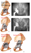
What is the classification of femoral head AVN?
[JAAOS 2014;22:455-464]
- Ficat and Arlet
- Stage 1 = normal
- Stage 2A = sclerotic or cystic lesions (no crescent sign)
- Stage 2B = sclerotic or cystic lesions with crescent sign (subchondral collapse without flattening of the femoral head)
- Stage 3 = flattening of the femoral head
- Stage 4 = osteoarthritis with decreased joint space and articular collapse
- Steinberg (University of Pennsylvania) classification (modification of Ficat by addition of MRI/bone scan)
- Stage 0 = normal radiograph + normal MRI/bone scan
- Stage 1 = normal radiograph + abnormal MRI/bone scan
- Stage 2 = lucent and sclerotic changes
- Stage 3 = subchondral collapse (crescent sign) of femoral head without flattening
- Stage 4 = flattening of the femoral head
- Stage 5 = joint narrowing and/or acetabular changes
- Stage 6 = advanced degenerative changes

What is the Crowe classification for hip dysplasia?
[J Am Acad Orthop Surg 2002;10:334-344]
- Four categories based on the extent of proximal migration of the femoral head
- Calculated on an AP pelvis xray by measuring the vertical distance between the inter-teardrop line and the junction between the femoral head and medial edge of the neck.
- The amount of subluxation is the ratio between this distance and the vertical diameter of the undeformed femoral head (gives the %)
- If both femoral heads are deformed the reference vertical height is the pelvic vertical height (highest point on the iliac crest to the inferior margin of the ischial tuberosity)
Type 1 – <50% proximal migration of the femoral head
- <10% of the vertical height of the pelvis
Type 2 – 50-75% proximal migration of the femoral head
- 10-15% of the vertical height of the pelvis
Type 3 – 75-100% proximal migration of the femoral head
- 15-20% of the vertical height of the pelvis
Type 4 - >100% proximal migration of the femoral head
- >20% of the vertical height of the pelvis

What is the Hartofilakidis classification?
[Orthop Clin N Am 43 (2012) 369–375]
- Describes acetabular abnormality rather than degree of subluxation
- Three categories:
1. Dysplastic hip- The femoral head is contained within the original acetabulum despite the degree of subluxation
- Acetabular deficiencies
- Segmental deficiency of the superior wall
- Secondary shallowness due to fossa covering osteophyte
- Low dislocation
- The femoral head articulates with a false acetabulum that partially covers the true acetabulum to a varying degree
- Acetabular deficiencies
- Complete absence of the superior wall
- Anterior and posterior segmental deficiency
- Narrow opening and inadequate depth of the true acetabulum
- High dislocation
- The femoral head is completely out of the true acetabulum and migrated superiorly and posteriorly to a varying degree
- Acetabular deficiencies
- Segmental deficiency of the entire with narrow opening
- Inadequate depth
- Excessive anteversion
- Abnormal distribution of bone stock, mainly located superoposteriorly

Described Acetabular Reconstruction in DDH
[J Am Acad Orthop Surg 2002;10:334-344]
Features of the acetabulum in DDH:
- Shallow, deficient anteriorly, laterally and superiorly, anteverted
- Goal when feasible is to place the acetabular component in the true acetabulum to restore the hip center
Crowe Type 1
- Standard acetabular component in the true acetabulum, medialize to inner table if needed to obtain coverage
- The acetabular component is usually small (38-52mm) with size usually being limited by the anterior and posterior dimensions of the native acetabular walls and distance between anterior and posterior columns
Crowe Type 2/3
- Options
- “Augmentation with bone graft”
- Place acetabular component in the true acetabulum with augmentation of the superolateral defect (autograft femoral head)
- Modular porous metal augments are also acceptable
- Place acetabular component in the true acetabulum with augmentation of the superolateral defect (autograft femoral head)
- “High hip centre”
- Place the acetabular component superior to the true acetabulum in the ‘high hip centre’
- “Medialization of the cup’
- Place the acetabular component in a medialized position with intentional overreaming or even fracture of the medial wall
- “Augmentation with bone graft”
Crowe 4
- Extra-small acetabular component in the true acetabulum without bone grafting

Femoral Reconstruction in DDH
[J Am Acad Orthop Surg 2002;10:334-344]
Features of the femur in DDH
- Excessive anteversion
- Coxa valga
- Small medullary canal
- Posteriorly located GT
Crowe 1/2
- Reconstruction with either uncemented or cemented (avoid excessive anteversion)
- Nonmodular, proximally porous-coated, metaphyseal-filling implants can be used when anatomic abnormalities are mild
- Extensively porous coated diaphyseal fitting implants bypass the proximal deformities and allow for adjustments in version (cannot be used when canal is very small)
- Modular implants allow use of proximally porous coated stem and adjust for proximal deformity
- Cemented implants allow adjustments in version
Crowe 3/4
- Femoral shortening is usually required
- Options:
- Combination of greater trochanteric osteotomy, sequential proximal femoral resection, and insertion of a cemented stem (technically easier)
- Subtrochanteric shortening osteotomy and insertion of a uncemented stem

What are the features of the periacetabular osteotomy?
[JAAOS 2011;19:275-286]
- Approach – abductor-sparing modification of the Smith-Petersen anterior iliofemoral approach
- 4 osteotomies
- Anterior ischium below the acetabulum; superior pubic ramus; supra-acetabular ilium; and posterior column, joining cut number one
- ***Note – posterior column is left intact
- Acetabular repositioning
* The acetabular fragment is tilted laterally, rotated inward, medialized, and extended (anteriorly tilted) - Acetabular fixation
* Achieved with three or four 4.5mm cortical screws - Arthrotomy performed
- Labral debridement or repair
- Osteochondroplasty at head neck junction allowing for increased hip flexion, internal rotation, and abduction
- Occasionally, in the presence of severe coxa valga, concomitant varus producing intertrochanteric femoral osteotomy is performed to optimize coverage

What is the definition of coxa profunda?
Deep hip socket where the medial wall of the acetabulum is medial to the ilioischial line (but the femoral head is not)

How do you assess for (THA) poly wear radiographically?
[JAAOS 2017;25:288-296]
Compare distance from superior rim of the acetabular component to superior aspect of femoral head with the distance from the inferior aspect of the femoral head to the inferior rim of the acetabular component (Compare A to B in image).

How do you assess for osteolysis on the acetabular and femoral sides based on radiographs?
[JAAOS 2017;25:288-296]
- Acetabular osteolysis – assess the 3 Charnley zones
- Femoral osteolysis – assess the 14 Gruen zones
***Use multiple plain radiograph views and serial xrays to assess progression

What are the technical aspects of cementing a liner into a well fixed acetabular component?
[JAAOS 2017;25:288-296]
- Prepare the acetabular component by using a high speed burr to score radial and circumferential grooves (~2mm in width and depth)
- If using a liner not designed for cementing it should be prepared with similar grooves on the backside
* Ensure the poly is of adequate thickness to allow for the grooves - The cement mantel should be ~2mm thick

What are the radiographic findings associated with pelvic discontinuity?
[JAAOS 2017;25:330-338]
- AP pelvis
- Visible fracture line
- Obturator ring asymmetry
- Medial migration of the inferior hemipelvis with disruption of Kohlers line
- Cross-table lateral
* Visible fracture line through posterior column - Judet views
* Fractures through both the anterior and posterior columns

What are the surgical options available to address pelvic discontinuity?
[JAAOS 2017;25:330-338]
- Posterior column compression plating and hemispheric acetabular component
- Should augment with 3-4 screws (ideally into ischium and superior ramus)
- Indications:
- Acute pelvic discontinuity
- Intraoperative discontinuity
- Chronic discontinuity and good bone stock
- Cup-cage construct
- Cup = Highly porous metal jumbo acetabular component (≥62mm in women and ≥66mm in men) +/- highly porous metal augments
- Cage = placed over the cup and spanning from ilium to ischium
- Cup and cage are unitized by a screw that passes through cage, cup and into host bone
- Polyethylene liner is cemented into the cup cage construct
- Indications – compromised bone quality or quantity (including irradiated bone)
- Distraction method
- Stability is achieved by distraction of the discontinuity and resulting elastic recoil of the pelvis
- Further stability is achieved with screws into the superior and inferior hemipelvis
- Healing occurs to the cup rather than across the fracture
- Technique involves sequential reaming until the anterosuperior and posteroinferior margins of the acetabulum are engaged
- A jumbo cup 6-8mm larger than the last reamer is used to obtain the distraction
- It is fixed in place by press fit supplemented by screws into the ilium and ischium
- A jumbo cup 6-8mm larger than the last reamer is used to obtain the distraction
- Indications – stiff discontinuity where modest distraction leads to elastic recoil
4. Custom triflange components - Custom designed porous and/or hydroxyapatite coated, titanium acetabular components with iliac, ischial, and pubic flanges
- Rigid fixation promotes healing of the discontinuity
- CT scan required for customization