Diseases Flashcards

1
Q

Causes for iron deficiency?

A

BLOOD LOSS #1

iron-poor diet

malabsorption

20% pre-menopausal women, 50% preggers

2
Q

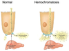
What is hemochromatosis?

A

genetic disorder, autosomal recessive

caused by mutation in HFE gene mostly

patients absorb excessive iron

iron deposits in liver, heart, pancreas, pituitary gland, other tissues

since liver affected, risk of cirrhosis and hepatocellular carcinoma

since pancreas affected - damage to islet cells -> DM in 2/3 of patients

increase in skin pigmentation as a result of increased melanin and increased iron deposition within the skin

cardiac deposition -> cardiac damage, CHF, arrhythmias

pituitary land iron -> drop in production of gonadotropins (sex hormones) -> testicular atrophy, etc

arthritis common - up to 1/2 of patients

takes years to develop serious disease - usually presents in mid 30ies-40ies

-> patients with hemochromatosis do not make enough hepscidin

3
Q

Acquired hemochromatosis?

A

iron overload can be acquired

first deposited into RE cells of the liver, spleen, bone marrow (R - connective tissue cells with reticulocytes - special form of collagen; RE cells = macrophages that break down RBCs)

as RE cells saturate, iron deposited in liver, pancreas, heart and other organs -> resembles hereditary

can be complication of anemias with ineffective erythropoiesis, such as beta-thalassemia - iron absorption increased and multiple blood transfusions add even more iron

alcoholics with liver disease can develop increased iron stores, since hepcidin made in the liver

increased iron consumption by itself does not cause hemochromatosis - body regulates absorption

4
Q

Causes of B12 deficiency?

A
  • poor nutrition (vegans)
  • pernicious anemia (ex. pariental cells damaged - no IF = intrinsic factor)
  • total or partial gastrectomy (since need IF from stomach to transport B12 to intestine for absorption)
  • intestinal disease (need intestinal factors to transport and absorb)
5
Q

Causes of folate deficiency?

A
  • poor nutrition (elderly, powerty, alcoholics)
  • increased utilization (pregnancy, lactation, malignancy - need for DNA synthesis, inflammation, etc)
  • intestinal disease (link to B12)
  • drug induced
6
Q

What is megaloblastic anemia (do not confuse with macrocytic anemia)?

A
  • B12 and folate deficiency most common causes of megaloblastic anemia, since DNA synthesis is defective
  • RBCs (erythroblasts) in bone marrow show delayed maturation of nucleus relative to cytoplasm - cells bigger b/c cytoplasm grows more before nucleus can mature
  • circulating RBCs are large (macrocytic) with marked variation in size and shape
  • megaloblastic anemia is one of the types of microcytic anemia
7
Q

Role of folate and B12 in blood?

A

B12 - cofactor for two biochem reactions

  1. homocysteine-> methione which generates tetrahydrofolate (THF) from methyl THF. need tetrahydrofolate because that is the active form of folate inside cells
  2. methylmalonyl coA -> Succinyl CoA (part of citric acid/Krebs - in the middle of the cycle)

Folate

cofactor in numerous biochem reactions

required to synthesize a precursor of DNA

both important for RBC maturation -> not enough -> DNA sythesis for RBC slows down, cytoplasm increases in relation to nucleus -> larger cells = macrocytic anemia

8
Q

List mechanisms for anemia (3)?

A

* blood loss

* decreased production

* increased destruction

9
Q

What is reticulocyte count?

A
  • reticulocyte count - marrow’s ability to produce young RBCs in response to turnover reticulocyte - immature RBC, 1% or so normal
  • increased reticulocyte count - adequate production of young RBCs to satisfy decreased RBC destruction (hemolysis, recovery post hemorrhage, therapy -ex. Fe-deficiency anemia and you give them iron)
  • decreased reticulocyte count - inadequate stores (Fe, B12, folate - slow RBC production) or to marrow abnormality (decreased ability to produce RBCs)
  • interpret with Hb
10
Q

Outline approach to diagnosing anemia based on RBC size?

A

Microcytic = decreased MCV ) ~ <82

Normocytic = normal MCV (mean corpuscular volume) 82-98

Macrocytic = increased MCV -100+

11
Q

List differentials for microcytic anemia?

A
  • small TAILS
  • Thalassemia (missing one of the Hb chains)
  • Anemia of chronic disease (b/c reduced iron release from RE macrophages - less iron available to be used for RBC synthesis) = also in malignancy and infection
  • Iron deficiency (b/c not enough building blocks for Hb)
  • Lead poisoning
  • Sideroblastic anemia
12
Q

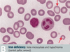
Discuss pathogenesis of iron deficiency anemia?

A
  • iron is part of Hb and rate-limiting for erythropoiesis

Causes (decreased intake, decreased absorption = increased elimination)

  • # 1 - GI (men) or uterine (women) bleeding
  • bleeding from GI tract
  • menorrhagia (heavy or prolonged menstrual period)
  • pregnancy
  • malabsorption (celiac, gastritis, GI malignancy, hookworm)
  • malnutrition
  • bleeding from GU
13
Q

Common symptoms of anemia?

A
  • SOB
  • fatigue
  • dizziness
  • weakness
  • pallor
  • palpitations - heart starts beating faster to maintain adequate O2 supply
  • tinnitus (ringing in ears)
  • pica (appetitne for non-food substances, like ice or dirt)
  • weak hair, nail flattening and concaven nails = koilonychia
  • conjunctival pallor
14
Q

Investigations for anemia?

A
  • # 1 CBC and peripheral blood smear
  • look for cause - consider endoscopy or colonoscopy if change in bowel habits or new indigestion
  • fecal occult blood test is useful to exclude GI bleed
  • can test urine for hematuria
  • chest X-ray to exclude very rare pulmonary haemosiderosis
  • sometimes no cause found (20%)
    *
15
Q

Results of investigations?

A
  • low ferritin is diagnostic of iron deficiency! (ferritin = storage form of iron, iron wrapped in proteins)
  • ferritin is also an acute phrase reactant iand is elevated in inflammatory condtions and liver disease
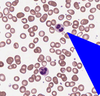
  • peripheral blood film shows hypochromic microcytosis - small RBCs with large white centre (hypochromic), can also have target cells
  • increased TIBC (iron wants to bind to transferrin more to be transported to tissues)
  • decreased serum ferum (iron bound to transferrin low, b/c low iron)
  • decreased iron saturation - less iron bound to transferrin b/c less iron in general
16
Q

Treatment for anemia?

A
  • treat underlying cause
  • supplement iron
    • oral - tablets/syrup - ferrous sulphate or ferrous gluconate (ferrous = Fe2+)
    • IV if cannot tolerate or absorb iron (iron sucrose or dextran)
  • monitor response - want increased reticulocyte count, increased Hb, will need to be on supplements for 4-6 months
17
Q

Pathogenesis of alpha thalassemia?

A

Defect in alpha globulin gene - decreased synthesis

4 alleles total for alpha globulin

if all 4 missing - no alpha globulin - incompatible with life - Hb Barts (gamma 4) - hydrops fetalis

3 allele deletion - HbH disease - very little alpha globulin - excess beta globin forms beta 4 Hb (HbH) - decreased MCV (mean cell volume), decreased Hb, splenomegaly (breakdown of malfunctioning alpha globulins)

1-2 allele deletion - virtually asymptomatic

alpha for A = Asia = South East Asia (SEA) and Africa

HbA 97% of adult hemoglobin

HbA2 - 3% of adult hemoglobin

both contain two alphas

18
Q

Pathogenesis for beta thalassemia?

A

Point mutation - decreased beta blobin synthesis

prevalent in mediterranean populations

beta -thalassemia minor (heterozygote) - less beta chains, usually asymptomatic

diagnosis confirmed by increased HbA2 (which has 2 alphas and 2 gammas (no betas) on electrophoresis

beta-thalassemia major (homozygote) - absent beta -> severe anemia, requiring blood transfusions -> can lead to secondary hemochromatosis ; here autosomal recessive

marrow expansion

increased HbF (2 alpha 2 gamma) - HbF naturally exists until 6 months, so infant only symptomatic after 6 months, when HbA takes over -> severe anemia and jaundice, then stunted growth and development, hepatosplenomegaly (extramedullary hematopoiesis - outside of bone marrow), expanded bone marrow - b/c of increased RBC synthesis

HbS (sickle) or beta thalassemia heterozygote - mild to moderate sickle sickle cell disease depending on amount of beta globulin

  • marrow expands in an effort to produce more RBCs (hemoglobin not as effective in transporting O2, so need more RBCs) -> bone enlargements (ex. ‘hair-on-end’ fluffy appearance on Xrays of skull) -> if untreated bone fractures, ulcers, etc
19
Q

Testing for alpha thalassemia and treatment?

A

Testing:

peripheral blood film - screen for HbH inclusion bodies with special stain

Hb electorphoresis not diagnostic for apha-thalasssemia - 4 copies of alpha

DNA analysis is the only way to confirm

**Treatment: **

depends on degree: if 1 or 2 defective alleles - no treatment required

if 3 (HbH disease) - transfuse

if 4 (HbBarts) - intrauterine transfusion (incompatible with life - not born otherwise)

20
Q

Testing and treatment for beta thalassemia?

A

Investigations:

CBC - low Hb

increased # RBCs - body tries to compensate for low Hb by making more RBCs -but RBCs smaller - missing Hb particles - microcytic anemia - also leads to bigger bones on Xrays (hematopoiesis is in bone marrow, breakdown in spleen - splenomegaly)

Hb electrophoresis: HbA really low (b/c beta parts missing), HbA2 high (b/c does not contain beta), HbF high (even though should be at 0 after 6 months

Treatment:

lifelong transfusions to stop endogenous production of Hb

iron chelation because increased transfusions lead to hemochromatosis

splenectomy - now less frequent

common in mediterranean patients

21
Q

Describe pathogenesis, tests and treatment for lead poisoning?

A

**Pathogenesis: **

lead inhibits enzymes involved in heme synthesis and increases RBC protoporphyrin (empty porphyrin without Fe), also lead inhibits rRNA degradation, which causes RBCs to have basophilic stippling (purple basophilic dots)

high risk in old houses with chipped paint

Test:

blood lead levels - consider occupational history

abdo pain, constipatino, irritability, lack of concentration

Treat: chelation therapy

LEAD

Lead Lines on gingivae (Burton lines) and on long bones on x-rays

Encephalopathy and erythrocyte basophilic stippling (purple dots)

Abdominal colic and sideroblastic Anemia

Drops - wrist and foot drop. Dimercaprol and eDta to chelate, Succimer in kids - “sucks to be a kid who eats lead

22
Q

Discuss pathogenesis of anemia of chronic disease?

A

Anemia of inflammatory, infective and malignant disesases (malignancy, RA, connective tissue disorders, chronic infections or extensive trauma)

  • inflammation -> IL6 released -> stimulates hepcidin release from liver -> hepcidin inhibits ferroportin (channel that pumps iron into body from enterocyte) -> reduced plasma levels
  • macrophage trapping iron -> reduced plasma levels
  • marrow unresponsive to normla or slightly elevated EPO - reduced RBC

rarely severe (Hb usually >90 vs 130 N in males, 120N in females)

very common - probably #2 after iron deficiency

inflammatory cytokines, such as IL-1 and IL-6 -> esp IL-6 can affect hepcidin (released from liver) directly, hepcidin binds to ferroportin and blocks absorption of iron from GI -> decreased plasma iron concentration

thought that maybe hits reduces Hb-O2 carrying capacity - limits O2 to microorganisms to help fight infection

anemia of chronic disease -> decreased iron levels - > decreased RBC production (also in part due to blunted response to erythropoietin and reduced red cell survival)

23
Q

Tests for anemia of chronic disease?

A
  • elevation in acute phase reactants (CRP, fibrinogen, ferritin)
  • serum iron and TIBC low b/c lower circulating iron, but % saturation normal, ferritin normal (iron stores not affected)
  • Hb slightly down
  • serum iron concentration and transferrin concentration are reduced (less iron)
  • serum ferritin level is normal or high - serum ferritin is acute phase reactant and increases in inflammation, malignancy and infection
  • normocytic (mild anemia of chronic disease) or sometimes microcytic (moderate anemia of chronic disease)
24
Q

Management of anemia of chronic disease?

A
  • manage underlying disorder
  • usually condition non-severe and non-progressive; treat only if Hb too low
  • IV iron if PO not working
  • if cannot compensate, consider blood transfusion, can give erythropoietin if chronic renal failure
25
Q

Summarize blood findings in microcytic anemias?

A
26
Q

What is sideroblastic anemia?

A

Sideroblasts are erythrocytes with iron-containing (basophilic) granules in the cytoplasm

“normal” iron-containing granules are small and randomly spread in the cytoplasm in healthy individuals.

“ring” : iron deposits in mitochondria, forming a ring around the nucleus - large granules in sideroblastic anemia - this is from bone marrow b/c peripheral blood would not show nucleus or ribosomes

“ring sideroblasts are found exclusively in pathological conditions, and should not be confused with ferritin sideroblasts, which are present in normal bone marrow. “

due to defect in heme synthesis - hereditary, X-linked or acquired (ex. myelodysplastic syndrome) or drug-related

27
Q

Investigations and treatment of sideroblastic anemia?

A

Investigations:

  • increased serum Fe2+, normal TIBC, increased ferritin (increased iron, which is what produces basophilic stippling)
  • ringed sideroblasts on peripheral blood smear (mitochondria with lots of iron around nucleus)
  • RBCs are hypochromic, basophilic stripping
  • can be micro, normo or macrocytic

Treatment:

depends on etiology

28
Q

Pathophysiology of sickle cell anemia?

A

part of hemoglobinopathies, primarily affects Afro-Carribean population (thought to be protective against malaria, since RBC changes shape and parasite cannot inhabit it)

betaSbeta S - serious disorder

betaSbetaThalassemia - serious, similar

betaSbetaNormal - sickle trait

betaS - one of the amino acids in Hb sequence replaced - in deoxygenated state HbS changes shape from flat tetramer to tetramers that pile on top of each other making a long polymer. cell looses its ability to restore shape and stays sickle shaped due to membrane damage -> sequestration of RBCs in microvasculature -> extreme pain and ischemia and increased hemolysis due to sequestration in RE network (like spleen)

due to this hemolysis, make sure patient on folate to prevent further drop in RBCs

29
Q

Symptoms and complications of sickle cell anemia?

A
  1. haemolytic anemia

RBCs do not retain normal shape - increased breakdown in the spleen, spleen can also be clogged by them and ischemic ->

  1. functional asplenism - surface of spleen decreases over time due to clogging by RBC and/or vascular ischemia -> susceptibility to infection increases! especially parvovirus B19
  2. vascular occlusive crises - acute, episodic painful crises triggered by infections, clod, etc -> MSK pain that is so severe it can require hospital admission. attacks often self-limiting but infarction can occur.

worry about strokes and transient ischemic attacks!

also responsible for “hand-foot syndrome” - dactylitis damaging small bones in hands and feet - swelling

  1. acute chest syndrome - 30% - dyspnea, chest pain, fever, tachypnea, leukocytosis, pulmonary infiltrate on CXR. caused by vaso-occlusion, infection or pulmonary fat embolus from infarcted marrow.
30
Q

Investigations for sickle cell anemia?

A
  • sickle cell prep - lower O2 so cells sickle “detects sickling of RBCs under microscope in response to O2 lowering agent”
  • Hb electrophoresis to distinguish btwn HbAS (trait), HbSS (homozygous), HbSC (one of S + thalassemias)
  • CBC - increased reticulocytes in homozygous and S+thal, decreased Hb
  • peripheral blood - sickled cells
  • electrophoresis - no HbA, only HbS and HbF, normal HbA2 (no beta there)
31
Q

Treatment for sickle cell anemia?

A
  • genetic counselling
  • folic acid to prevent folic deficiency (want as many RBCs produced as possible, folic acid limiting reactant)
  • hydroxyurea to enhance production of HbF (b/c no beta there)
  • if vaso-occlusive crisis: give O2, liquid to reduce viscosity, correct acidosis if present (since acidosis or high CO2 can also precipitate), analgesia for pain, transfuse if very serious - acute chest or stroke, multi-organ failure, ICU
  • avoid hypoxia, acidosis, dehydration, fever - anothing that leads to sickling, vaccinate in childhood b/c of functinal asplenism
  • screen for complications (bloodwork inc CBC, BUN, creatinine), urinalysis for proteinuria and glomerulopathy, transcranial doppler for stroke prevention, retinal exam from 8 yo for retinopathy, echo from 10 yrs for pulmonary hypertension (remember acute chest common)
32
Q

What is aplastic anemia?

A

Caused by failure or destruction of myeloid stem cells due to :

* radiation and drugs

* viral agents like parvovirus B19, EBV, HIV, HCV

* Falconi anemia (DNA repair defect)

* idiopathic

=> pancytopenia and hypocellular bone marrow (less cells, emptyish bone marrow)

33
Q

Symptoms of aplastic anemia?

A

Symptoms

* general anemic symptoms (b/c reduced RBCs)

* thrombocytopenia and/or infection from pancytopenia

* splenomegaly and lymphadenopathy may be present

34
Q

Investigations for aplastic anemia?

A

* exclude other causes

* CBC - anemia or neutropenia or thrombocytopenia in any combination (if anemia, tired, if neutropenia infection-prone, if thrombocytopenia, increased bleeding)

* decreased reticulocytes (<1% of the total RBC count (think life 120 days, so 120/100% = 1% of new ones per day)

* blood film = peripheral blood smear = decreased number of RBCs

* bone marrow - decrease in marrow cells with fat replacing them

35
Q

Treatment for Aplastic Anemia?

A
  • remove offending agents (if drugs, etc)
  • supportive care - RBC/platelet transfusions, antibiotics - careful b/c transfusions can increase immune sensitization to blood products
  • allogenic bone marrow transplant
36
Q

What are causes for macrocytic anemia?

MCV > 100 fL

A

ABCDEF

  • Alcoholism (liver disease)
  • B12 deficiency
  • Compensatory reticulocytosis
  • Drugs/Dysplasia
  • Endocrine (hypothyroidism)
  • Folate deficiency/Fetus (pregnancy)

macrocytic anemia can be megaloblastic and non-megaloblastic

megaloblastic would have alrge, oval nucleated RBC precursors due to failure of DNA synthesis - cytoplasm matures longer than normal b/c it takes a while for the nucleus to form

in non-megaloblastic anemia, cells are abnormal size because of membrane abnormality or abnormal cholesterol metabolism

37
Q

Discuss Vitamin B12 deficiency anemia?

A

B 12 = cobalamin, hypersegmented neutrophils

B12 binds ot R protein to pass through stomach, than pancreatic enzymes digest that complex in small intestine and B12 binds to IF - intrinsic factor made in stomach by parietal cells. Only when B12 is attached to IF can it be absorbed by the body (terminal ileum)

Deficiency:

decreased intake (vegan, vegetarian in pregnancy)

decreased absorption:

gastric trouble - mucosal atrophy, pernicious anemia, post-gastrectomy (decreased IF production)

malabsorption due to Crohn’s, celiac sprue, pancreatic insufficiency, tapeworm, resection of ileum (site of absorption), drugs

B12 important because converts dietary folate into usable form, which can then participate in DNA synthesis (no B12, no usable folate - no DNA synthesis - large cells, b/c nucleus takes forever to sythesize, meanwhile cytoplasm grows extreme -> megaloblastic anemia). B12 also participates in Kreb’s cycle ??? (double check last bit)

38
Q

What is the pathophysiology of pernicious anemia?

A
  • auto-antibodies produced against gastric parietal cells leading to lack of intrinsic factor secretion
  • IF cannot bind to B12 -> B12 cannot be aborbed by the body

Get:

neurological symptoms!!!! b/c B12 participates in fatty acid pathways and myelin synthesis

cerebral symptoms (reversible with B12 therapy) - confusion, delirium, demetntia

cranial nerve symptoms (optic atrophy, etc)

peripheral neuropahty - lower limbs affected more

39
Q

Investigations in pernicious anemia? Treatment?

A
  • CBC - MCV > 110 fL, low reticulocyte count, neutropenia an dthrombocytopenia can be present
  • serum B12 and RBC folate low
  • oval macrocytes, hypersegmented neutrophils on peripheral blood smear = blood film
  • elevated unconjugated bilirubin and LDH due to breakdown of cells in BM
  • Schilling test
  • Schilling test: Step 1 - givve B12 PO, and a dose of unlabeled B12 to saturate tissues so radiolabled B12 excreted in urine, measure how much excreted - if normal excretion, shortness of B12 due to dietary deficiency, its absorption/excretion normal
  • Schilling test: Step 2: give radiolabeled B12 with intrinsic factor (oral IF) if first stage shows reduced excretion
  • if normal test - pernicious anemia (shortness of IF, fixed when IF added)
  • if abnormal test - intestinal causes, like malabsorption (not diet or IF production, something not absorbing past IF stage)

Treat by supplementing B12 IM montly for life, or PO daily

40
Q

What is Schilling test? describe 2 steps

A

test to understand why B12 short:

Schilling test: Step 1 - givve B12 PO, and a dose of unlabeled B12 to saturate tissues so radiolabled B12 excreted in urine, measure how much excreted - if normal excretion, shortness of B12 due to dietary deficiency, its absorption/excretion normal

Schilling test: Step 2: give radiolabeled B12 with intrinsic factor (oral IF) if first stage shows reduced excretion
if normal test - pernicious anemia (shortness of IF, fixed when IF added)
if abnormal test - intestinal causes, like malabsorption (not diet or IF production, something not absorbing past IF stage)

41
Q

Discuss folate deficiency? (all)

A

uncommon in developed countries due to dietary supplementation - in greens, and leafy vegetables

causes: malnutrition (alcoholics), malabsorption, antifolate drugs (methotrexate,etc), increased requirements, like in hemolytic anemia, pregnancy

no neurological manifestations

similar findings to B12 deficiency, rule out B12 deficiency as cause

give folic acid PO - but understand that it will mask B12 deficiency and neurological degeneration will continue if thats the cause so rule it out first

42
Q

List differentials for normocytic anemia?

A
  • sickle cell disease
  • aplastic anemia
  • acute blood loss
  • hemolysis
  • anemia of chronic disease (microcytic or normocytic)
43
Q

What are key features of MDS (myelodysplastic syndrome)?

A

Suspect MDS (myelodysplastic syndrome) if:

  • elderly patient
  • history of chemo
  • gradual onset
  • macrocytic anemia
  • pancytopenia
  • hypolobated neutrophils = 2 lobes vs 3-5N “pseudo-Pelger-Huet neutrophils “

ex. case 78 yo female Hx progressive fatigue, no FHx, clear Abdo, Chest, Cardiac and rectal exams (to rule out blood per rectum), no hematuria, epistaxis (nose bleeds). breast cancer at age 65 (breast exam also negative). no weight loss, fevers, etc - to rule out malignancy.

44
Q

Medical name for low WBC count?

A

leukopenia

(normal 4-11X10 9/L - provided)

45
Q

Medical name for low Hb count?

A

anemia

normal : over 110 in pregnant 120 in women 130 g/L in men

46
Q

Medical name for low platelet count?

A

thrombocytopenia

normal range 150-400X10 9 /L

acute phase reactant

47
Q

Name division for macrocytic anemias?

A

Megaloblastic

(B12, folate deficiencies) - rare

Non-megaloblastic

  • drugs,
  • liver disease (participates in breakdown of heme, etc),
  • hypothyroidism (see card) - thyroid hormones stimulate erythropoiesis
  • myelodysplastic syndromes,
  • marrow failure syndromes
48
Q

Why does hypothoroidism contribute to anemias?

A

Thyroid hormones generally stimulate erythropoiesis. These agents also increase erythrocyte 2,3-DPG concentrations, which serve to enhance the delivery of oxygen to tissues.

Anemia is an uncommon finding in hyperthyroidism but when present may be morphologically similar to that observed in hypothyroidism.

49
Q
A
50
Q

What is G6PD deficiency?

A

extravascular or intravascular

common enzymatic disorder, X-linked recessive

Defect in G6PD -> decrease in glutathione -> RBCs much more susceptible to oxidant stress

ex. Hb oxidizes -> denatures and precipitates - > damage to RBC membrane

Causes include: antimalarials (oxidize), fava =broad beans (high in oxidative components), infections, sulfa drugs

51
Q

What are Heinz bodies?

A

Hb gets oxidized and precipitates in G6PD deficiency

Some of it gets removed from RBCs directly by spleen - > bite cells and blister cells

Some of it looks like it precipitated and RBCs have dots -> Heinz bodies (also in alpha thalassemia)

(Oxidative) stress makes me eat bites of fava beans iwth Heinz ketchup

52
Q

What are clinical features of G6DP deficiency?

A
  • X-linked recessive
  • episodes of hemolysis after
    • oxidative stress
    • drugs (sulfas, antimalarials, etc)
    • infection
    • food (fava beans high in oxidative components)
    • in neonates can present as jaundice
      *
53
Q

Investigations, results and treatment for G6PD?

A
  • G6PD assay available
  • blood film will show Heintz bodies and bite cells (b/c spleen bites off abnormally precipitated Hb (b/c of oxidation) off RBCs, whereas Heintz bodies are cells that are not bitten off with Hb precipitated
  • avoid triggers as treatment, transfusion if very severe

When in (oxidative) stress, I bite into fava beans with Heintz ketchup.

54
Q

What is pyruvate kinase deficiency?

A
  • autosomal recessive, rare
  • defect in pyruvate kinase -> glucose cannot break down (enzyme for one of last steps) -> decrease in ATP -> rigid RBCs with increased hemolysis
  • hemolytic anemia in the newborn
  • extravascular hemolysis
55
Q

What is HbC defect?

A
  • extravascular lysis
  • point mutation on beta-globin - one amino acid changed
  • patients with HbSC (1 of each mutant gene) have a milder disease than HbSS patienst (sickle cell)
56
Q

What is hereditary spherocytosis?

A
  • defect in vertical membrane proteins of RBCs (band 3, protein 4.2, ankyrin, spectrin)
  • less membrane -> small and round RBCs with no central pallor - > premature RBC removal by spleen
  • spleen makes defective RBCs even more spherocytotic and fragile by removing their membrane faster since fragile - each round in spleen - less membrane more spherical cells
  • extravascular lysis
  • Labs - osmotically fragile (when put in salt solution burts a lot faster than normal RBCs (b/c already round and full of fluid), spehorcytes
  • in severe cases splenectomy and vaccinations against infections (pneumococcus, meningococcus and H.influenza (since prone to infections without spleen)
  • MCHC - mean cell Hb. concentration up - cells pack more content,
  • RDW - distribution width up b/c sizes vary with spherocytosis
  • treat with splenectomy
57
Q

What is hereditary elliptosis?

A
  • abnormal RBC membrane interactions (horizontals), like spectrin alpha/beta, protein 4.1 etc
  • autosomal dominant
  • hemolysis usually mild
  • elliptical RBCs on blood smear
  • immunize b/c spleen to busy removing cells (immune function decreased), splenectomy in very severe hemolysis
58
Q

For membrane disorder questions, what is the membrane structure of RBCs?

A
59
Q

Causes of normocytic anemia:

A

as simple as ABCDs

**A - **acute blood loss (GI, etc)

**B - **bone marrow failure = aplastic anemia = destruction of blood cells in the marrow

**C- ** chronic disease

**D - **destruction - hemolysis

  • membranous (genetic) spherocytosis and elliptocytosis due to mutations in membrane proteins on RBC
  • enzymatic (genetic): G6PD deficiency (enzyme that fights oxygenation of Hb and free radicals), pyruvate kinase deficiency - enzyme that is important in glucolysis (less ATP in RBCs)
  • Hb: thalassemias - abnormal shape -lysis; hemoglobinopathies
  • acquired: hemolytic transfusion reactions, autoimmune hemolytic anemia, drugs, etc

s - Sickle cell anemia

Or think SAHARA

S- sickle cell

A- aplastic anemia (bone marrow trouble)

H - hemolytic anemia (sphero/elliptocytosis, G6PD, pyrivate kinase deficiency, thalassemias (increase destruction), etc

A - anemia of chronic disease

R - nothing really

A - acute loss of blood

  • Simplification:*
    normocytic: loss of RBCs - acute blood loss;

malformed RBCs that get destroyed

  • (spherocytosis, eliptocytosis, pyruvate kinase and G6PD deficiency, thalassemias)
  • sickle cell
  • aplastic anemias (problem with total formation in bone marrow)

normal RBCs that get destroyed (hemolytic transfusion, autoimmune, anemia of chronic disease)

60
Q

Some symptoms of normocytic anemia?

A

See:

jaundice (increased destruction -> increased bilirubin)

dark urine (b/c more Hb, bilirubin - bilirubin gives urine colour)

cholelithiasis (b/c increased bilirubin)

aplastic crisis possible (if severe lack of blood cells, as in aplastic anemia, overwhelming infection can happen (lack of WBCs, neutrophils, etc)

61
Q

What is hemolytic uremic syndrome, how does it present?

A

Ex. E.coli (“hamburger disease”) - hemorrhagic reaction if O157: H7 -> secretes toxin which is absorbed by colon, toxin carried in blood stream -> kills enterocytes (bleed from GI) and endothelial lining in small vessels, like in kidneys -> kidneys damaged, high urea and creatinine, anemia due to increased bleeding (but normocytic) - > since endothelial cells in kidney damaged, body lays down fibrin clots to prevent bleeding in these vessels (increased D-dimer, which is a measure of clots in the body!) -> as RBCs pass these narrow vessels, they can damaged -> schistocytes on smear

Renal failure is due to the injury to small endothelial vessels in the kidney caused by E. coli toxin (from EHEC type) -> renal vasculature damaged -> loss of kidney function -> hemolytic uremic syndrome

62
Q

Differential for microcytic anemias?

A

Since small, think SMALL TAILS

TAILS

thalassemias

anemia of chronic disease (can be normocytic too)

iron deficiency

lead

sideroblastic anemia

63
Q

What is sideroblastic anemia?

A

Sideroblastic anemia - uncommon

  • erythrocytes with iron-containing (basophilic) granules in the cytoplasm
  • these granules are present normally too, but then they are small and spread out in healthy individuals
  • in sideroblastic anemia, there is a “ring” around the nucleus of them, as iron deposits in mitochondria, abnormal, large granules vs small ones
  • can be X-linked (rare), drug-driven, etc
  • investigations: increased serum Fe2+, normal TIBC, increased ferritin
  • get ringed sideroblasts, basophilic stippling
64
Q

What is HFE gene? What conditions does it play in?

A

HFE gene - gene that encodes human hemochromatosis protein. this protein functions to regulate iron absorption by regulating interaction between *transferrin receptor* with transferrin (transferrin is protein that TRANSports Ferritin (iron) in the blood)

hereditary hemochromatosis (HHC) is an auto recessive disorder usualy from defects in this gene -> excess iron storage

common - 1:200 people of Northern European descent are carriers

65
Q

Mechanisms for thrombocytopenia?

A
  • reduced production
  • sequestration (ex. in spleen)
  • increased destruction/comsumption
66
Q

What are some examples of reduced platelet production?

A

Ex .genetic like Bernard Soulier syndrome can be associated with decreased platelet production (Bernard Soulier - platelet adhesion to vWF and consequently sheared vessel (GpIbIX) -> inability of primary hemostasis b/c platelet cannot cover bleed (fibrin is secondary hemostasis)

Ex. acquired like B12/folate deficiency - cannot synthesize

infiltrative - hematologic/ non hematologic cancers

marrow failure like myelodysplastic syndromes (abnormal myeloid lineage blood cell development - past blast stage but immature - most destroyed before leaving bone marrow - cytopenias), aplastic anemias

medications, like chemo

Platelet transfusions effective but should be reserved for serious bleeds/surgery

67
Q

What are some examples of sequestration leading to thrombocytopenias?

A
  • spleen sequestration - up to 1/3 of platelets
  • if splenomegaly, this # increased ex: congestive like in liver cirrhosis/portal HTN
    • spleen also reactive to infections, autoimmune, benign or malignant
  • transfusions don’t help much (more sequestration as we add more platelets
68
Q

What are some examples of increased consumption of platelets leading to thrombocytopenia?

A
  • immune thrombocytopenia = idiopathic thrombocytopenia = immune thrombocytopenic purpura
  • drug induced thrombocytopenia (like heparin induced HIT)
  • sepsis
  • DIC - disseminated intravascular coagulation
    *
69
Q

What is immune thrombocytopenic purpura = immune thrombocytopenia (ITP) = idiopathic thrombocytopenia?

A
  • most often anti GpIIb/IIIa antibodies (ones that help platelets aggregate with fibrin) -> splenic macrophages (reticuloendocytes) kill platelet antibody comples -> decreased platelet life
  • may be triggered by viral illness
  • chronic and acute ITP
  • children manage condition well and recover quickly (more often acute, 1 week after viral illness), may be chronic in adults
  • can be triggered by meds like sulfa, associated with rheum conditions since autoimmunish
  • lab MAY show increased megakaryocytes on bone marrow biopsy (megakaryocytes are precursors to platelets (up to 1:4000), try to restore platelet number); thrombocytopenia
  • decreased platelet count, consequently increased bleed time
  • treat with steroids (since autoimmune), can also give immunoglobulins (to fight antibodies), do splenectomy in severe cases (since spleen destroys these platelets)
  • platelet transfusion not effective - > platelets get destroyed again
70
Q

What are some secondary causes of thrombocytopenias (ex. HIT, etc?)

A

Drug-induced thrombocytopenia: drug complexes with platelets -> complex antibodies are produced. Withdraw offending drug (substitute with another class), transfuse platelets if bleeding is significant.

Heparin Induced Thrombocytopenia (HIT) develops 4-14 days post-treatment – often thrombocytopenia accompanied with micro thromboemboli (complexes aggregates and clog arteries).
PF4 protein in alpha granules of platelets -> binds to circulating heparin -> PF4 –heparin complex immunogenic -> PF4-heparin-IgG complex -> binds platelet surfaces -> causes platelet activation, degranulation (granule secretion) and platelet aggregation -> severe thromboembolic predisposition and consumptive thrombocytopenia

1-5% of patients, thrombocytopenia if \<100 platelets
**stop heparin and give a different class of anticoagulant**

Post-transfusion purpura – 1 week post blood transfusion, treat with IVIg (immunoglobins)

71
Q

What is DIC (disseminated intravascular coagulopathy)?

A

Disseminated intravascular coagulation (DIC), also known as disseminated intravascular coagulopathy, characterized by the widespread activation of the clotting cascade that results in the formation of blood clots in the small blood vessels throughout the body. This leads to compromise of tissue blood flow and can ultimately lead to multiple organ damage. In addition, as the coagulation process consumes clotting factors and platelets, normal clotting is disrupted (b/c everything used up) -> severe bleeding can occur from various sites. DIC does not occur by itself but only as a complicating factor from another underlying condition (sepsis, malignancy,etc) usually in those with a critical illness. The combination of widespread tissue ischemia and simultaneous bleeding carry an increased risk of death in addition to that posed by the underlying disease. DIC can be overt and severe in some cases, but milder and insidious in others.

May give platelet transfusion

72
Q

Some additional things that can cause coagulopathies?

A

Congenital Disorders of Platelet Function ~ Qualitative platelet disorders

Bernard-Soulierautosomal recessive, rare – mutation of Gp (glycoprotein) IbIX – responsible for adhesion of platelet and von Willebrand factor to subendothelium (through small microfibers exposed). Bernard-Soulier leads to platelet dysfunction, thrombocytopenia and abnormal morphology. DDAVP (to release factor VIII from stores, which in turn is a carrier for von Willebrand) helps.

Glanzmann’s thrombastheniaautosomal recessive, rare – mutation of Gp IIb/IIIa, which links fibrinogen and platelets. Platelet number normal, but they cannot aggregate. Usually neonatal onset, mucous and GIbleeds, menorrhagia (heavy periods).

Others: lack of granules in platelets:

lack of alpha granules = alpha granule shortage (proteins for aggregation and repair of broken endothelium) or dense granule shortage (Ca++, ADP/ATP, serotonin)

defective granule content release – inhibits platelet activation => aspirin like effect, since ADP encourages platelet aggregation and aspirin blocks AA -> COX1-> TXA2 which also encourages platelet aggregation.

Acquired Disorders of Platelet Function ~ Qualitative platelet disorders

Drugs:

Aspirin – irreversibly activates cyclooxygenase-1 (enzyme) = COX 1 -> prevents production of TXA2 (thromboxane A2) from AA -> impaired platelet activation and aggregation for platelet life span (7-10 days) -> prolongs bleeding time

Clopidogrel inhibits ADP binding to receptor -> ADP mediates platelet aggregation - > impaired (irreversible for life of platelet (7-10 days)

Heparin binds platelets; can inhibit aggregation

IIbIIIa inhibitors: inhibit platelet aggregation (binds fibrinogen to platelets)

Systemic conditions:

Chronic renal failure = uremia (accumulation in the blood of toxic wastes that are supposed to be eliminated by kidneys, present in severe kidney disease)

~ elevated nevels of NO inhibit platelet adhesion, activation, aggregation

Cardiopulmonary bypass – platelets are activated and degranulated in the extracorporeal circuit (outside of body), reducing their effectiveness in vivo. Excessive bleeding uncommon, platelet transfusion can help.

Haematologic disease (too many platelets, too few or proteins impair aggregation)

Myelodysplastic syndromes (the blood stem cells (immature cells) do not become healthy red blood cells, white blood cells, or platelets -> blasts formed (immature) and die in either bone marrow or soon after release in blood -> # of healthy blood cells reduced -> infections, anemia, easy bleeding)

Myeloproliferative syndromes (abnormal growth of RBCs, WBCs and platelets in the bone marrow)– platelet dysfunction may occur especially with very high platelet counts

73
Q

What is Thrombotic Thrombocytopenic Purpura (TTP)?

A

Thrombotic Thrombocytopenic Purpura is a group of microvascular occlusive disorders - systemic or intrarenal aggregation of platelets, thrombocytopenia and mechanical injury of erythrocytes.

Triggered by:

  • toxin-mediated endothelial cell damage (hemolytic uremic syndrome with Shiga-like toxin damaging endothelial cells - hypercoagulation and renal damage)
  • congenital absence of metalloproteases (enzymes) that cleave vWFactor called ADAMTS13 (scary stuff) - familial TTP
  • autoantibody causing deficiency of ADAMT13 (primary TTP)
    • in both of latter ones won Villaebrand factor is cleaved into a large multimer - giant vWF attracts platelets in large doses - microcoagulation
74
Q

What is the classic pentad for Thrombotic Thrombocytopenic Purpura?

A
  • thrombocytopenia
  • fever
  • neurologic symptoms
  • renal impairment (think HUS)
  • microangiopathic hemolytic anemia (think HUS - damage to RBCs as blood vessels occluded with partial clots)

=> accumulation of large wWF multimers - > huge platelet aggregation -> microthrombi, mortality of over 90% if untreated (10% if treated)

75
Q

Treatment for thrombotic thrombocytopenic purpura?

A
  • medical emergency
  • plasma exchange - to remove autoantibody (if acquired) and large vWF multimers
  • replacement of plasma provides fresh source of metalloprotease enzymes that will cut vWF normally
  • aspirin often added to reduce microthrombi
  • PLATELET TRANSFUSIONS COUNTERINDICATED - > fear of more coagulation with more platelets
76
Q

Discuss polycythemia vera?

A

One of chronic myeloproliferative disorders, polycythemia = increased hematocrit

often non-hereditary mutation in JAK2 gene, which binds to EPO receptors and stimulates them even with low EPO -> excessive hematocrit production.

can present with intense itching (b/c of increased histamine production), aslo blood clotting, redness, etc

77
Q

What is essential thrombocytosis?

A

Similar to polycythemia vera, but specific to overproduction of abnormal platelets. Platelets can function well -> increased thrombosis or abnormallly - > increased bleeding.

Bone marrow will contain enlarged megalkaryocytes (precursors to platelets)

78
Q

What is myelofibrosis?

A

part of chronic myeloproliferative disordrs - fibrotic obliteration of bone marrow - bone marrow replaced with fibrotic cells. Teardrop RBCs and immature forms of the myeloid line cells (bone marrow crying b/c it is fibrosed)